Educational Blog about Anesthesia, Intensive care and Pain management

Showing posts with label Congenital cardiovascular diseases. Show all posts

Ebstein’s Anomaly

Ebstein’s Anomaly


-A rare congenital cardiac abnormality.

-The septal and posterior cusps of the tricuspid valve are displaced downwards and are elongated, such that a varying amount of the right ventricle effectively forms part of the atrium. Its wall is thin and it contracts poorly. The remaining functional part of the right ventricle is therefore small.

-The foramen ovale is patent, or defective, in 80% of cases.

-The degree of abnormality of right ventricular function, and the size of the ASD, are probably the main determinants of the severity of the condition, which varies considerably.

-The right ventricular systolic pressure is low, and the RVEDP is elevated. Tricuspid incompetence can occur.

-There may be a right to left shunt, with cyanosis, on effort, and pulmonary hypertension and right heart failure may supervene.

-The natural history of the disease is very variable. Fifty percent of cases present in infancy with cyanosis, and 42% die in the first 6 weeks of life.

-In those who survive to adulthood, symptoms may be precipitated by the onset of arrhythmias, or by pregnancy. A few patients remain asymptomatic, even as adults, although once symptoms develop, the disability can increase rapidly.

-A cardiothoracic ratio of ≥ 0.65 is a better predictor of sudden death than the symptomatic state, and those who developed atrial fibrillation died within 5 years. It has therefore been suggested that tricuspid surgery should be undertaken before the cardiothoracic ratio reaches 0.65.

Preoperative Abnormalities:

1. There may be a right to left shunt, with dyspnea and cyanosis at rest, or on moderate exertion. Alternatively, the patient may be asymptomatic.

2. Episodes of tachyarrhythmias occur in 25% of patients. Some provoke syncopal attacks.

3. The ECG may show varying abnormalities, including large peaked P waves, a long P–R interval, Wolff–Parkinson–White syndrome, RBBB, and right heart strain. Paroxysmal supraventricular tachycardia occurs in 15%, usually because of the presence of WPW syndrome.

4. Chest X-ray may show cardiomegaly, with a prominent right heart border, and poorly perfused lung fields.

5. Paradoxical systemic embolism and bacterial endocarditis may occur.

6. Many other lesions of the tricuspid valve or right ventricle may mimic Ebstein’s anomaly, therefore the discriminating clinical and echocardiographic features for correct diagnosis have been enumerated.

Anesthetic Problems:

These will depend upon the anatomical abnormality, the degree of right to left shunt, and the presence or absence of right heart failure.

1. Induction time is prolonged, because of the pooling of drugs in the large atrial chamber.

2. Intracardiac catheter insertion may be hazardous because it can provoke serious cardiac arrhythmias.

3. Air entering peripheral venous lines or any open veins at subatmospheric pressure may cause paradoxical air emboli.

4. Tachycardia is poorly tolerated because of impaired filling of the functionally small right ventricle.

5. Hypotension may increase the right to left shunt if present.

6. Hypoxia causes pulmonary vasoconstriction, which also increases a right to left shunt.

7. There is a risk of bacterial endocarditis, especially if a CVP line is in place.

8. Deterioration may occur in pregnancy because of a decrease in right ventricular function, and an increase in blood volume and cardiac output, or with the onset of arrhythmias.

Management:

1. The severity of the lesion must be assessed. In the presence of maternal cyanosis or arrhythmias during pregnancy, there should be close monitoring of both mother and fetus. Deterioration may occur, despite previous successful pregnancies.

2. Treatment of heart failure and arrhythmias.

3. Antibiotic prophylaxis against bacterial endocarditis.

4. If a CVP is used for monitoring, its tip should be kept within the superior vena cava. The use of intracardiac catheters should probably be avoided.

5. Techniques should aim to minimize tachycardia and hypotension.

6. Oxygen therapy increases pulmonary vasodilatation. Long-term maternal therapy is required during pregnancy from 14 weeks, to treat fetal hypoxia that is demonstrated by umbilical venous blood gases.

7. Several anesthetic techniques have been described. A two-catheter epidural technique can be used for vaginal delivery to minimize hypotension. Bupivacaine doses must be fractionated and saline rather than air used to site the epidural, to avoid paradoxical air emboli. Cesarean section under general anesthesia, preceded by fentanyl, and a neurolept analgesic technique for hysterectomy, have been described.

Eisenmenger’s Syndrome

 Eisenmenger’s Syndrome

Eisenmenger’s Syndrome


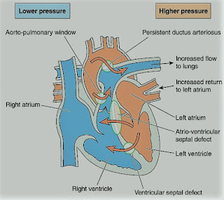
-A rare syndrome of pulmonary hypertension associated with a reversed or bidirectional cardiac shunt, occurring through a large communication between the left and right sides of the heart. The defect may be interventricular, interatrial, or aortopulmonary.

-The development of Eisenmenger’s syndrome, from the initial left to right shunt, is usually a gradual process. Contributory factors to pulmonary hypertension are hypoxia, high pulmonary blood flow, and high left atrial pressure.

-Irreversible structural changes take place in the small vessels, causing pulmonary vascular obstruction and a reduction in the size of the capillary bed. The pulmonary artery pressure is the same as or sometimes exceeds, the systemic arterial pressure.

-The incidence of this syndrome is decreasing because of the more vigorous approach to diagnosis and treatment of congenital heart disease in childhood.

Preoperative Findings:

1. Presenting symptoms include dyspnea, tiredness, episodes of cyanosis, syncope, or chest pain. Hemoptysis may occur.

2. The direction of the shunt, and hence the presence or absence of cyanosis, depends on several factors. These include hypoxemia, pulmonary and systemic pressure differences, and intravascular volume. It can also be affected by certain drugs.

3. Sleep studies have shown that there is a nocturnal deterioration in arterial oxygen saturation, which seems to be related to ventilation/perfusion distribution abnormalities occurring in the supine position.

4. Chest X-ray shows right ventricular hypertrophy, and ECG indicates varying degrees of right ventricular hypertrophy and strain.

5. Complications include thrombosis secondary to polycythemia, air embolus, bacterial endocarditis, gout, cholelithiasis, and hypertrophic osteoarthropathy. A cerebral abscess may occur secondary to clot embolism.

Anesthetic Problems:

1. Reductions in systemic arterial pressure by myocardial depression or loss of sympathetic tone are potentially dangerous. Reversal of the shunt may occur, and sudden death has been reported.

Hypovolemia and dehydration are poorly tolerated. Syntocinon may cause a dramatic reduction in SpO2 secondary to vasodilatation.

2. Sinus tachycardia results from exercise or emotion, and episodes of SVT are common after the age of 30. The onset of atrial fibrillation is associated with a sudden deterioration in the condition of the patient.

3. General anesthesia tends to be favored since the reduction in systemic vascular resistance associated with regional blockade increases the shunt. However, successful use of epidural anesthesia for bilateral inguinal herniorrhaphy, and Cesarean section, have been reported.

4. Pregnancy is contraindicated because it carries considerable risks. Recent maternal mortality rates of 40% have been reported. A cesarean section may increase it to over 60%. Termination of pregnancy is usually recommended in the first trimester but is still associated with a mortality of 7%.

5. Patients are at risk from paradoxical air or clot embolism, and infective endocarditis.

Anesthetic Management:

1. Understanding the pathophysiology of the complex is essential, and both pregnancy and non-cardiac surgery require a multidisciplinary approach.

2. Maintenance of an adequate circulating blood volume is important. Myocardial depressants and peripheral vasodilators should be used with caution. Bradycardia must be prevented. If regional anesthesia is used, the block should be instituted with caution, and hypovolemia avoided.

3. It is unclear whether oxygen can cause pulmonary vasodilatation. Although the pulmonary vascular resistance was believed to be fixed in pulmonary hypertension, a high oxygen concentration has been shown to reduce it during cesarean section.

4. Maintenance of systemic vascular resistance is critical. The use of a norepinephrine infusion before induction has been described. Alpha-adrenergic vasopressors, such as methoxamine or phenylephrine, have also been used for the treatment of hypotension on induction of anesthesia.

5. Pulmonary ventilation should be performed with low inflation pressures and early tracheal extubation is advised, because of the deleterious effects of IPPV.

6. Air must be completely eliminated from all intravenous lines and the epidural space should be located with loss of resistance to saline, not to air.

7. Antibiotic prophylaxis against bacterial endocarditis.

8. Low-dose heparin may reduce the risk of emboli.

9. Patients are usually advised against pregnancy. If anesthesia is required either for termination of pregnancy or operative delivery, intensive cardiac care is indicated. It has been suggested that those reaching the end of the second trimester should be admitted to the hospital until delivery and given heparin 20000-40000 units daily and oxygen therapy. Successful epidural anesthesia has been reported for cesarean section.

Tetralogy of Fallot

 Tetralogy of Fallot

Tetralogy of Fallot


-Tetralogy of Fallot (TOF) is a congenital cardiac abnormality. The primary defects are pulmonary infundibular stenosis and ventricular septal defect (VSD). The VSD is sufficiently large for the pressure in both ventricles to be equal to that of the aorta. The tetralogy is completed by two secondary features, a variable degree of overriding of the aorta, and right ventricular hypertrophy.

-Dynamic right ventricular outflow obstruction may occur (infundibular spasm), which is accentuated by sympathetic stimulation. The fraction of the right to left shunt depends primarily upon the relative resistances between the pulmonary (or right ventricular) and systemic outflows.

-If TOF is associated with patent foramen ovale (PFO) or atrial septal defect (ASD), it is a Pentalogy of Fallot.

-The aim of the surgery is to relieve the right ventricular outflow obstruction and to close the VSD. The traditional management by a two-stage repair has been replaced by definitive correction. Recent surgical advances include conduits to connect the RV to the PA and transatrial repair of the VSD.

The problems encountered during anesthesia will depend upon whether or not corrective surgery has been undertaken, and the functional result.

Preoperative Findings:

1. Dyspnea may occur on exertion and is hypoxia-related. Cyanosis and finger clubbing are variable, depending on the degree of pulmonary stenosis and the size of the shunt. Polycythemia (erythrocytosis) is common. There is a pulmonary stenotic murmur, but no murmur from the VSD because of the size of the defect. Squatting is thought to reduce the fraction of the shunt since kinking the large arteries increases systemic vascular resistance. Squatting is commonly seen in children with uncorrected lesions.

2. ECG shows right atrial and right ventricular hypertrophy, right axis deviation, and right bundle branch block.

3. Echocardiography shows right atrial and right ventricular hypertrophy, VSD, PFO or ASD, pulmonary hypertension, and overriding of the aorta.

4. Chest X-ray shows right ventricular hypertrophy and oligemic lungs. In the 2.6–6% of individuals who also have an absent pulmonary valve, aneurysmal dilatation of the pulmonary arteries may cause bronchial compression.

5. Initial surgery may have been undertaken to anastomose a systemic to a pulmonary artery, to improve the pulmonary blood flow, and reduce cyanosis. A definitive procedure is now more commonly undertaken in infancy.

6. In patients who have undergone shunt surgery without a definitive repair, there is chronic hypoxia and polycythemia. These patients have a high mortality, and an increased risk of bacterial endocarditis, thrombotic stroke, emboli, and intracerebral abscess.

7. In adults who have undergone repair there is an increased risk of arrhythmias, conduction defects, and sudden deaths, possibly related to mechanical events, such as ventricular dilatation and stretch, in the proximity of the conduction system. However, new approaches to surgery may result in a lower incidence of such problems in the future.

Anesthetic Problems:

1. In individuals with uncorrected lesions, the right to left shunt, and hence the cyanosis, is increased by a reduction in systemic vascular resistance produced by systemic vasodilatation. This may result from factors such as hypovolemia, drugs effect, or pyrexia.

2. Cyanosis is also worsened by an increase in pulmonary vascular resistance or spasm of the right ventricular infundibulum. Right ventricular outflow obstruction is due to increased contractility which is produced by increases in catecholamine output or the administration of drugs with positive inotropic effects. Anxiety, pain, hypercarbia, hypoxia, and acidosis are all precipitating factors. These cyanotic attacks or ‘tet’ spells, which can occur when awake or under anesthesia, may initiate a cycle of increasing hypoxia that can result in cerebral damage or death. Direct intraoperative observations of shunt direction and flow have been made with Doppler color flow imaging using epicardial leads. Patients with severe life-threatening hypoxemic spells, refractory to other treatment, responded to phenylephrine (5 mcg/kg plus an infusion of 0.4–2 mcg/kg/min).

3. Dehydration in the presence of polycythemia and high plasma viscosity may combine to increase the incidence of cerebral thrombosis. Polycythemia may also be associated with coagulation defects.

4. In patients with an absent pulmonary valve, positional airway compromise occurred secondary to bronchial compression of dilated pulmonary arteries.

5. A significant incidence of tracheal anomalies has been found.

6. In adults who have undergone repair, ventricular and atrial arrhythmias are common, particularly during exercise. It has been suggested that patients scheduled for elective surgery should have Holter monitors, or undergo exercise testing, in case antiarrhythmic treatment is needed first.

7. Cyanosed patients with Fallot’s rarely become pregnant. However, adults who have undergone corrective surgery are increasingly present during pregnancy with a favorable outcome.

Anesthetic Management:

1. Antibiotic prophylaxis against bacterial endocarditis.

2. A good premedication (morphine sulphate, or midazolam) to prevent excitement and anxiety.

3. In patients with cyanosis, measures are aimed at reducing the right to left shunt. Specific treatments of cyanotic attacks include:

a) Oxygen 100% to decrease PVR.

b) Pressor agents, such as phenylephrine (5-10 mcg/kg), to increase systemic vascular resistance.

c) Fluids to correct hypovolemia.

d) Propranolol (0.1 mg/kg) or Esmolol (0.5 mg/kg) to decrease outflow tract obstruction (decrease contractility and infundibular spasm).

e) Deepening of light anesthesia to reduce tachycardia associated with catecholamine output.

f) Compression of the femoral artery or the abdominal aorta against the vertebrae, sufficiently firmly to stop the femoral artery pulsations.

4. Techniques to avoid hypoxia and hypercarbia, and minimize vasodilatation and sudden increases in cardiac output. Ketamine, and Morphine sulphate (0.05-0.1 mg/kg), have been used.

5. Hydration is maintained in the perioperative period and, if there is severe polycythemia, venesection may be necessary.

6. Metabolic acidosis should be prevented or treated with sodium bicarbonate (1-2 mmol/kg).

7. In pregnancy, the outcome is satisfactory in patients whose ventricular function is good, and in whom no residual shunt occurs. However, close observation should be undertaken by an experienced team.

8. Venesection for erythrocytosis that is associated with cyanotic congenital heart disease should only take place if there are symptoms of hyperviscosity with a hematocrit >65%, and only provided volume replacement takes place at the same time.

9. Avoid muscle relaxants with histamine release e.g. atracurium, and use cisatracurium or rocuronium.

9. Mechanical ventilation:

-Increase FiO2

-Avoid increased airway pressure (< 15 cmH2O)

-Increase respiratory rate

-Avoid PEEP

Coarctation of Aorta

 Coarctation of Aorta

Coarctation of Aorta


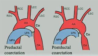
-A congenital narrowing of the aorta that may be pre-or post-ductal.

-The preductal form is usually a long, narrow segment, and is associated with other cardiac defects. This type generally presents with heart failure before the age of 1 year and requires treatment in a pediatric cardiac surgical unit.

-The postductal form, however, is often asymptomatic, and the patient may present in later life for surgery of some other condition, or for correction of the coarctation itself. Even after correction of coarctation, abnormalities can continue.

-Those who have undergone repair show persistent alterations in left ventricular function and left ventricular mass, together with resting gradients between the arm and leg. There is a higher incidence than normal of ischemic heart disease and sudden death.

-The optimal time for repair is disputed. Hypertension is more common in late correction, but repair at a young age is associated with a higher risk of recoarctation. In addition, persistent abnormalities in aortic stiffness have been found.

-Adult repairs are associated with a higher incidence of significant aortic valve disease (58%), compared with 37% of those repaired in childhood.

Preoperative Findings:

1. There may be moderate hypertension, the arm blood pressure being higher than that in the leg. If the left subclavian arises at or below the constriction there may be an absent or reduced left radial pulse. If both radial and femoral pulses are felt together, the small volume and delay of the femoral pulse will be obvious.

2. Collateral circulation develops in the internal mammary, intercostal, and subscapular arteries. The latter may be seen if the scapula is illuminated from the side.

3. A systolic murmur is usually heard along the left sternal edge radiating up into the neck.

4. Chest X-ray may show notching of the undersides of the ribs, secondary to intercostal arteries dilatation. There may be pre-and post-stenotic dilatation of the aorta.

5. Occasionally, cerebral berry aneurysms coexist with coarctation. In such cases, the high arterial pressure increases the risk of subarachnoid hemorrhage.

6. There is a 25–50% incidence of the bicuspid aortic valve and some aortic regurgitation.

7. Unusually, angina or left ventricular failure may present late in untreated adult coarctation.

8. Patients may require balloon dilatation or surgical resection.

Anesthetic Problems:

1. Before elective surgery, if undiagnosed coarctation is found, treatment of the coarctation may be considered to be the priority. Even if coarctation has been treated, the possibility of residual cardiovascular abnormalities should be considered, since there is an increased risk of premature death compared with the normal population. Causes include aneurysms (cerebral, at the operative site, other parts of the aorta and intercostal arteries), hypertension, myocardial infarction, and cardiac failure. Balloon angioplasty may carry less risk.

2. Patients may present with hypertension-related complications.

3. Any operation in the area of the dilated collateral vessels may result in heavy bleeding, especially when the chest is opened.

4. Hypoperfusion of the spinal cord. This may cause paraplegia, and is more likely in those patients with few collaterals. Induced hypotension for clipping of cerebral aneurysms may compromise spinal cord perfusion.

5. Susceptibility to bacterial endocarditis.

6. If there are left subclavian abnormalities, the left arm cannot be used for blood pressure monitoring.

7. Patients may present during pregnancy with hypertension, which may be confused with preeclampsia.

Anesthetic Management:

1. Antihypertensive therapy should be used until the day of operation. Beta-adrenoceptor blockers may reduce the hypertensive response to intubation.

2. Antibiotic prophylaxis against bacterial endocarditis.

3. During clipping of an intracranial aneurysm, monitoring of the femoral artery pressure should be undertaken to assess spinal cord perfusion during induced hypotension. A mean distal aortic pressure above 50 mmHg has been suggested as adequate for spinal cord perfusion.