Educational Blog about Anesthesia, Intensive care and Pain management

Showing posts with label ICU. Show all posts

Ventilators

 Ventilators


-Early ventilators consisted of the generation of negative pressure around the whole of the patient’s body except the head and neck; these were called Cabinet or Iron lung ventilators.

-A negative pressure could also be applied over the thorax and abdomen: Cuirass Ventilators.

Classification:

1. Pattern of gas flow during inspiration:

a) Pressure generators:

Constant pressure is produced by bellows or a moderate weight which produces a decreasing inspiratory flow which alters with changes in lung compliance

b) Flow generators:

Constant flow is produced by a piston, heavyweight, or compressed gas. Flow is unaltered by changes in lung compliance although pressures will vary. These ventilators have a high internal resistance to protect the patient from high working pressures.

2. Power:

Pressure generators are low powered whereas flow generators are high powered.

3. Cycling:

Change from inspiration to expiration may be determined by:

a) Time:

Most common method. The duration of inspiration is predetermined, with the constant flow it may be necessary to preset a Tv; when this has been delivered there is an inspiratory pause (improves distribution) before the inspiratory cycle ends.

b) Pressure:

Used as a pressure limit on other modes. Ventilator cycles into expiration when preset airway pressure is reached (delivers a different Tv if compliance or resistance changes). Inspiratory time varies according to compliance and resistance.

c) Volume:

Usually used with an inspiratory flow restrictor. Cycles into expiration whenever a preset Tv is reached.

d) Flow:

Older ventilators.

4. Sophistication:

Newer ventilators can function in many of the above modes, and also have weaning modes such as SIMV, PS, and CPAP.

5. Function:

a) Minute volume dividers:

Fresh gas flow powers the ventilator. Minute volume equals the FGF divided into pre-set tidal volumes, thus determining the frequency.

b) Bag squeezers:

Replaces the hand ventilation of a Mapleson D or circle system. It needs an external power source.

c) Lightweight portable:

Powered by compressed gas and consists of the control unit and patient valve.

Complications of Total Parenteral Nutrition (TPN)

Complications of Total Parenteral Nutrition (TPN)

Complications of Total Parenteral Nutrition (TPN)


I. Catheter-Related Complications:

-The hyperosmolarity of the dextrose and amino acid solutions requires infusion through large veins or central venous lines.

1-Misdirected Catheter: e.g., with subclavian vein cannulations – (mostly on the right) 10% resulted in misplacement of the catheter in the internal jugular vein.

2-Infection

3-Hematoma

4-Thrombosis

II. Carbohydrate Complications:

1-Hyperglycemia:

-Hyperglycemia is common during TPN; blood glucose levels >300 mg/dl were recorded in 20% of postoperative patients receiving TPN.

-A standard TPN regimen with 1,800 non-protein calories has ≈350 g of glucose, compared to 230 g in a standard tube feeding regimen.

-Tight glycemic control is not recommended in critically ill patients because of the risk of hypoglycemia, which has more serious consequences than hyperglycemia.

-The current recommendation for hospitalized patients is a target range of 140–180 mg% for blood glucose.

2-Insulin:

-If insulin therapy is required, regular insulin is preferred for critically ill patients, to prevent wide swings in glucose levels, by adding insulin to the TPN solutions.

-One shortcoming of IV insulin infusions is the propensity for insulin to adsorb to the plastic tubing in IV infusion sets. This affects the bioavailability of insulin but can be reduced by priming the IV infusion set with an insulin solution (e.g., 20 mL of saline containing 1unit/mL of regular insulin). But the priming procedure must be repeated each time the IV infusion set is changed.

-SC insulin can be used for stable patients. Regimens will vary in each patient, with a combination of intermediate or long-acting insulin with rapid-acting insulin, when needed.

3-Hypophosphatemia:

-The movement of glucose into cells is associated with a similar movement of phosphate into cells, and this provides phosphate for co-factors (e.g., thiamine pyrophosphate) that participate in glucose metabolism. This intracellular shift of phosphate can result in hypophosphatemia.

4-Hypokalemia:

-Glucose movement into cells is also accompanied by an intracellular shift of potassium (which is the basis for the use of glucose and insulin to treat severe hyperkalemia). This effect is usually transient, but continued glucose loading during TPN can lead to persistent hypokalemia.

5-Hypercapnia:

-Excess carbohydrate intake promotes CO2 retention in patients with respiratory insufficiency. This was originally attributed to the high respiratory quotient (VCO2/VO2) associated with carbohydrate metabolism. However, CO2 retention is a consequence of overfeeding, and not overfeeding with carbohydrates.

III. Lipid Complications:

-Overfeeding with lipids may contribute to hepatic steatosis.

-Triggering inflammatory response: The lipid emulsions used in TPN regimens are rich in oxidizable lipids, and the oxidation of infused lipids will trigger an inflammatory response. (Oleic acid, one of the lipids in TPN, is a standard method for producing ARDS in animals), and this might explain why lipid infusions are associated with impaired oxygenation.

IV. Hepatobiliary Complications:

1-Hepatic Steatosis:

-Fat accumulation in the liver (hepatic steatosis) is common in patients receiving long-term TPN and is believed to be the result of chronic overfeeding with carbohydrates and lipids. Although this condition is associated with elevated liver enzymes, it may not be a pathological entity.

2-Cholestasis:

-The absence of lipids in the proximal small bowel prevents cholecystokinin-mediated contraction of the gallbladder. This results in bile stasis and the accumulation of sludge in the gallbladder and can lead to acalculous cholecystitis.

V. Bowel Sepsis:

-The absence of nutritional bulk in the GI tract leads to atrophic changes in the bowel mucosa and impairs bowel-associated immunity, and these changes can lead to the systemic spread of enteric pathogens.

Bronchoscopy in ICU

Bronchoscopy in ICU


Indications:

A) Diagnostic:

1-Investigation of an infectious process:

➧ Bronchoscopy and broncho-alveolar lavage (BAL) are commonly used for the identification of pathogens in the lower airways in the following circumstances: 

a) When it is not clear whether the presence of organisms represents infection or colonization. 

b) When the infection is caused by organisms that show a predilection for the peripheral airways (e.g. Pneumocystis jirovesii). 

c) When the infection is confined in a particular area of the lung (tuberculous and non-tuberculous mycobacteria as well as several fungal and opportunistic organisms).

2-Abnormal breath sounds:

➧ Abnormal breath sounds include: stridor, wheezes, or a combination of both and they are the most common indication of FOB in the NICU.

➧ In neonates, abnormal breath sounds are due to congenital abnormalities of the upper or lower airways.

➧ In older infants and children the causes are often iatrogenic (e.g. subglottic stenosis due to prolonged intubation, injury of the vocal cords, or of the recurrent laryngeal nerve causing paresis or paralysis).

3-Evaluation of the nature of abnormalities:

➧ Patients in the ICU often present with radiographic findings and/or symptoms of unclear etiology, e.g.: an airway filled with secretions can look radiographically identical to a completely compressed airway.

4-Others:

➧ Pneumonia, Trauma, Inhalation injury and burns, Tracheoesophageal fistula.

B) Therapeutic:

1-Placement of the endotracheal tube:

➧ The use of FOB for the placement of the endotracheal tube is reserved for cases in which high precision is required.

➧ e.g. placement of the ETT just above the carina in patients with very severe tracheomalacia; or selective intubation of one lung), or when congenital anatomical abnormalities or injuries preclude the proper opening of the jaw for direct laryngoscopy.

2-Placement of double-lumen ETT and confirmation of position

3-Extubation over FOB

4-Persistent or recurrent atelectasis

➧ Atelectasis is a major cause of clinical deterioration and/or of delay in the patients’ recovery, resulting from many causes such as; mucous plug, compression of the airways, alveolar destruction, and collapse.

5-Foreign body removal

6-Strictures and stenosis

7-Hemoptysis

Contraindications:

1-Full stomach


3-Severe pulmonary hypertension

4-Tuberculosis

5-Acute myocardial infarction or unstable angina

6-Coagulopathy

Complications:

1-Increase HR, Bl. P, ICP, IOP

2-Laryngospasm and bronchospasm

3-Hypoxemia

4-Arrhythmia

5-Bleeding

6-Post-bronchoscopy fever

7-Pulmonary infiltrate

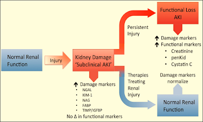
Acute Kidney Injury Biomarkers

Acute Kidney Injury Biomarkers

I) Functional markers

1-Serum Creatinine (SCr):

➧ It is a degradation product of muscle cells and represents a surrogate for the efficiency of glomerular filtration.

➧ It has poor predictive accuracy for renal injury, particularly, in the early stages of AKI.

➧ In the case of critical illness, SCr concentrations are subject to large fluctuations due to a patient’s induced dilutional volume status, the catabolic effects of critical illness, the likelihood of concentration decreases in septic conditions, and the increased tubular excretion with diminishing the renal function.

➧ Furthermore, after an injurious event, the rise in SCr is slow.

➧ Therefore, detection of the earliest evidence of AKI necessitates the use of other plasma or urinary biomarkers.

2-Plasma/Serum Cystatin C (CyC):

➧ It is a 13-kDa, non-glycosylated, cysteine protease inhibitor produced by all nucleated cells at a constant rate.

➧ In healthy subjects, plasma CyC (pCyC) is excreted through glomerular filtration and metabolized completely by the proximal tubules. There is no evident tubular secretion (not detectable in urine in healthy subjects).

➧ Several studies claim the superiority of pCyC against SCr to detect minor reductions in glomerular filtration rate (GFR).

➧ It is detected in plasma and urine 12-24 h. post-renal injury.

Confounding factors: older age, Gender, Weight, Height, Systemic inflammation, High levels of C-reactive protein, Malignancy, Thyroid disorders, immunosuppressive therapy, Glucocorticoid deficiency or excess, and Smoking.

3-Fractional Excretion of sodium (FENa):

➧ (FENa) measures the percent of filtered sodium that is excreted in the urine.

➧ This calculation is widely used to help differentiate prerenal disease (decreased renal perfusion) from acute tubular necrosis (ATN) as the cause of AKI.

➧ In pre-renal azotemia, the proximal tubules reabsorb filtered sodium resulting in a very low urine sodium concentration (<20 mmol/L) and FENa is <1%.

➧ In intrinsic AKI the urine sodium concentration is >40 mmol/L and FENa is >1%.

4- Proenkephalin A (Penkid):

➧ Penkid is a 5-kDa, stable breakdown product of enkephalins.

➧ It accumulates in the blood in settings of reduced GFR.

➧ It is associated with AKI and mortality in patients with sepsis and heart failure.

II) Low-molecular-weight proteins

1-Urine Cystatin C (uCyC):

➧ The urinary excretion of CyC (uCyC) specifically reflects tubular damage because systemically produced cystatin C is normally not found in urine.

2-Urine α1/β2 microglobulin:

III) Up-regulated proteins

1-Kidney Injury Molecule-1 (KIM-1) (Cytoprotection):

➧ It is a type I transmembrane glycoprotein with a cleavable ectodomain (90-kDa).

➧ It is localized in the apical membrane of dilated tubules in an acute and chronic injury.

➧ It is produced by proximal tubular cells after ischemic or nephrotoxic injury; no systemic source.

➧ KIM-1 plays a role in regeneration processes after epithelial injury and in the removal of dead cells in the tubular lumen through phagocytosis.

➧ A reduction in proteinuria with renin-angiotensin-aldosterone blockade is accompanied by a reduction in urinary KIM-1 excretion.

➧ It is detected in urine 12-24 h. after renal injury

Confounding factors: Renal cell carcinoma, Chronic proteinuria, Chronic kidney disease, Sickle cell nephropathy.

2-Neutrophil Gelatinase-Associated Lipocalin (NGAL) (also known as oncogene 24p3) (Iron binding):

➧ It is a 25-kDa glycoprotein produced by epithelial tissues throughout the body.

➧ It is a small protein linked to neutrophil gelatinase in specific leukocyte granules.

➧ It is also expressed in a variety of epithelial tissues associated with anti-microbial defense.

➧ NGAL’s composite molecule binds ferric siderophores, induces epithelial growth, has protective effects in ischemia, and is up-regulated by systemic bacterial infections.

➧ Plasma NGAL is excreted via glomerular filtration and undergoes complete reabsorption in healthy tubular cells. It is also produced in distal tubular segments.

➧ It is detected in plasma and urine 2-4 h. after AKI.

Confounding factors: Malignancy, Chronic kidney disease, Pancreatitis, COPD, Endometrial hyperplasia.

3-Liver Fatty Acid Binding Protein (L-FABP):

➧ They are small (15-kDa) cytoplasmic proteins (intracellular lipid chaperones) produced in tissues with active fatty acid metabolism (liver, intestine, pancreas, lung, nervous system, stomach, and proximal tubular cells).

➧ Their primary function is the facilitation of long-chain fatty acid transport, the regulation of gene expression, and the reduction of oxidative stress.

➧ Urinary (L-FABP) is undetectable in healthy control urine, which is explained by efficient proximal tubular internalization via megalin-mediated endocytosis.

➧ Under ischemic conditions, tubular L-FABP gene expression is induced. L-FABP is freely filtered in glomeruli and reabsorbed in proximal tubular cells; increasing urinary excretion after tubular cell damage.

➧ In renal disease, the proximal tubular re-absorption of L-FABP is reduced.

➧ Detected in plasma and urine 1 h. after ischemic tubular injury.

Confounding factors: Chronic kidney disease, Polycystic kidney disease, Liver disease, Sepsis.

4-Interleukin-18 (IL-18):

➧ It is 18-kDa pro-inflammatory cytokine, released from proximal tubular cells following injury.

➧ Detected in plasma and urine 6-24 h. after renal injury

Confounding factors: Inflammation, Heart failure, Sepsis.

5-Tissue Inhibitor of Metallo-Proteinases-2 (TIMP-2):

6-Insulin-like Growth Factor Binding Protein-7 (IGFBP-7):

➧ They are cell cycle arrest proteins that have been suggested as early indicators of AKI.

➧ In particular, urinary (TIMP-2) and (IGFBP-7) are biomarkers of the G1 renal tubular cell cycle arrest at the early phase of AKI.

➧ The product of the urinary concentrations of TIMP-2 and IGFBP-7 (urinary [TIMP-2] × [IGFBP-7]) is a promising biomarker for the early prediction of AKI.

IV) Tubular enzymes

1-Alpha-Glutathione-s-Transferase (α-GST):

2-Pi-Glutathione-s-Transferase (π-GST):

➧ (α-GST) and (π-GST) are 47-to 51-kDa cytoplasmic enzymes.

➧ They are both members of a multigene family of detoxification enzymes present in many organs including the kidney.

➧ Distribution across the entire nephron of structurally and functionally distinct isoforms has been demonstrated.

➧ In urine, these enzymes are normally not present.

➧ After the injury, α-GST is primarily detected in the proximal cells, whereas π-GST is observed in the distal parts.

➧ They are detected in urine 12 h. after AKI.

3-Gamma-Glutamyl Transpeptidase (GGT):

4-Alkaline Phosphatase (AP):

5-Alanine Amino-Peptidase (AAP):

➧ They are tubular brush border enzymes.

➧ They are released into the urine when there has been significant damage to the brush border membrane with loss of the microvillus structures.

6-N-Acetyl-β-D-Glucosaminidase (NAG):

➧ N-Acetyl-β-D-Glucosaminidase (NAG) is a lysosomal enzyme (>130-kDa) that is localized in the renal tubules.

➧ It precludes glomerular filtration (due to large MW), implying that urinary elevations have a tubular origin.

➧ Increased activity suggests injury to its cells but may also reflect increased lysosomal activity without cell disruption.

➧ NAG catalyses the hydrolysis of terminal glucose residues in glycoproteins.

➧ It is detected in plasma and urine 12 h. after AKI

Confounding factors: Diabetic nephropathy.

V) Others

1-Retinol Binding Protein (RBP):

➧ It is 21-kDa single-chain glycoprotein; a specific carrier for retinol in the blood (delivers retinol from the liver to peripheral tissues).

➧ It is totally filtered by the glomeruli and reabsorbed but not secreted by proximal tubules; a minor decrease in tubular function leads to excretion of RBP in urine.

➧ It is detected in plasma and urine

Confounding factors: Type II DM, Obesity, Acute critical illness.

2-Hepcidin:

➧ It is a 2.78-kDa peptide hormone predominantly produced in hepatocytes; some production in the kidney, heart, and brain.

➧ It is freely filtered with significant tubular uptake and catabolism (fractional excretion 2%).

➧ It is detected in plasma and urine after AKI.

Confounding factors: Systemic inflammation.

3-Hepatocyte Growth Factor (HGF):

➧ It is overexpressed after AKI.

➧ It is a marker linked to renal tubular epithelial cell regeneration.

4-Netrin-1:

➧ It is a laminin-related molecule, minimally expressed in proximal tubular epithelial cells of normal kidneys.

➧ It is highly expressed in injured proximal tubules.

➧ It is detected in urine after AKI.

5-Monocyte Chemo-attractant Peptide-1 (MCP-1):

➧ It is a peptide expressed in renal mesangial cells and podocytes.

➧ It is detected in urine after AKI.

Confounding factors: Variety of primary renal diseases.

6-Calprotectin:

➧ The calcium-binding complex of two proteins of the S100 group (S100A8/ S100A9).

➧ Derived from neutrophils and monocytes.

➧ Acts as an activator of the innate immune system.

➧ It is a measure of local inflammatory activity. It is detected in urine in intrinsic AKI.

Confounding factors: Inflammatory bowel disease, Urinary tract infection, Probably CKD.

7-MicroRNA:

➧ MicroRNAs are short, non-protein-coding RNA molecules between 19 and 25 nucleotides in length.

➧ They are epigenetic regulators of gene expression at the post-transcriptional level in response to kidney injury through messenger RNA (mRNA) signal repression.

➧ As key regulators of homeostasis, their dysregulation underlies several morbidities including kidney disease.

➧ MicroRNAs are used as diagnostic and prognostic biomarkers in AKI.

8- Chitinase-3-like protein 1 (CHI3L1) (YKL-40, HC-gp39):

➧ YKL-40 is a 40-kDa heparin- and chitin-binding glycoprotein also known as Human Cartilage glycoprotein 39 (HC-gp39), 38-kDa heparin-binding glycoprotein or chitinase-3-like protein 1 (CHI3L1).

➧ The abbreviation YKL-40 is based on the one-letter code for the first three N-terminal amino acids, tyrosine (Y), lysine (K), and leucine (L), and the apparent molecular weight of YKL-40.

➧ It plays an important role in AKI and repair.


Acute Kidney Injury Biomarkers

Acute Kidney Injury Biomarkers

Acute Kidney Injury Biomarkers