Educational Blog about Anesthesia, Intensive care and Pain management

Porphyric Crisis

Porphyric Crisis (Acute Neurovisceral Crisis) 

Background: 

-The porphyrias are caused by enzyme deficiencies in the heme production pathway. Such deficiencies may be due to inborn errors of metabolism or exposure to environmental toxins or infectious agents.

-The disease was named porphyria due to the red discoloration of urine in affected patients, (figure 1).

-Crises are four to five times more common in women and usually occur in their early 30s.

Porphyria red urine
Figure 1: Porphyria Red Urine

Triggering factors: 

Enzyme-inducing drugs: 

-Barbiturates (Thiopental, Methohexital), Etomidate, Enflurane, Alcuronium, Mepivacaine, Pentazocine, Nifedipine, Verapamil, Diltiazem, Phenytoin, Hydralazine, Phenoxybenzamine, Aminophylline 

Physiological: 

-Menstruation, Fasting, Dehydration, Stress, Infection, Anemia, Endogenous hormones 

Habits: 

-Smoking, Alcohol 

Clinical Picture: 

CNS: 

-Autonomic neuropathy (Fever, Pain, Constipation, Gastroparesis, Postural hypotension) 

-Peripheral neuropathy (Skeletal muscle weakness, Quadriparesis, Bulbar palsy, Respiratory failure) 

-Cranial nerve palsy 

-Seizures 

-Psychiatric features (Mood disturbance, Confusion, Psychosis) 

CVS: 

-Tachycardia, Hypertension 

GIT: 

Acute abdominal pain, Vomiting, Diarrhea, Dehydration, Electrolyte disturbance (↓ [Na⁺, K⁺, Mg⁺²]) 

Management: 

Remove triggering factors (above) 

Specific R: 

-Hematin, Heme arginate / Heme albumin, Somatostatin, Plasmapheresis 

Symptomatic R: 

-Pain: Substantial doses of Opioids 

-Nausea and Vomiting: Prochlorperazine, Ondansetron 

-Anxiety: Lorazepam, Midazolam in low doses 

-Insomnia: Zopiclone 

-Delirium: Haloperidol 

-Tachycardia & Hypertension: ÎČ-adrenergic blocking agents, Glyceryl trinitrate 

-Seizures: Benzodiazepines, Levetiracetam, Clonazepam, Gabapentin, Vigabatrin, Magnesium sulphate 

-Sedation: Propofol, Alfentanil infusions. The clinical safety of prolonged midazolam infusion is unknown. 

-Thromboembolic prophylaxis: LMW heparins 

-Stress ulcer prevention: IV Omeprazole, Ranitidine 

-Correction of electrolytes

Mechanical Ventilation for Respiratory failure

Carcinoid Crisis

Carcinoid Crisis

Carcinoid Crisis

Definition:

-Carcinoid crisis is the most serious and life-threatening complication of carcinoid syndrome and is generally found in people who already have carcinoid syndrome.

-Carcinoid crisis occurs when all of the symptoms of carcinoid syndrome come at the same time. 

Causes:

➧ Spontaneous 

➧ Precipitating factors: 

-Stress, Sympathetic stimulation 

-Hypotension 

-Histamine releasing drugs 

-Tumour manipulation 

-Regional anesthesia due to hypotension 

-Infection 

-Chemotherapy 

Clinical picture:

-Tachycardia, Arrhythmia 

-Hypotension, Shock 

-Flushing, Hyperthermia 

-Bronchospasm 

-Abdominal pain, Diarrhea 

Management:

Inhibit growth h. & vasoactive peptides release:

-Somatostatin analogs (Octreotide, Lanreotide) 

Anti-serotonin:

-Methesergide, Ketanserin, Cyproheptadine, Ondansetron, Alpha-methyl dopa 

Anti-kallikrein:

-Corticosteroids, Aprotinin

Anti-histamine:

-H1 blockers (Diphenhydramine) & H2 blockers (Ranitidine) 

R of Bronchospasm:

-Salbutamol, Aminophylline 

R of Diarrhea:

-Loperamide 

R of Hypotension:

-Vasopressin, Phenylephrine 

R of Hypertension:

-Alpha-blockers, Beta-blockers 

R of Rt. Heart failure:

-Digitalis, Diuretics

Sodium Nitroprusside Toxicity

Sodium Nitroprusside Toxicity:


Mechanism of Action:

After parenteral injection, sodium nitroprusside enters red blood cells, where it receives an electron from the iron (Fe⁺²) of oxyhemoglobin. This non-enzymatic electron transfer results in unstable nitroprusside radical and methemoglobin (Hb Fe⁺³). The former moiety spontaneously decomposes into five cyanide ions and the active nitroso (NO) group. 

The cyanide ions can be involved in one of three possible reactions: 

1) Binding to methemoglobin to form cyan-methemoglobin.

2) Undergoing a reaction in the liver and kidney catalyzed by rhodanase enzyme to form thiocyanate + thiosulfate.

3) Binding to tissue cytochrome oxidase, which interferes with normal oxygen utilization.

N.B. Sodium nitroprusside toxicity is usually related to prolonged administration or occurs in patients with renal or hepatic failure. 

Mechanisms of Toxicity:

1) Direct vasodilation: resulting in hypotension and dysrhythmias (most common).

2) Thiocyanate toxicity: (occurs infrequently).

3) Cyanide toxicity: (in rare cases).

4) Methemoglobinemia: (in very rare cases).

Thiocyanate toxicity :

Symptoms:

Anorexia, nausea, abdominal pain, fatigue, and mental status changes, including psychosis, weakness, seizures, tinnitus, and hyperreflexia. 

Treatment: 

-Toxicity can be minimized by avoiding prolonged administration of nitroprusside and by limiting drug use in patients with renal insufficiency (as thiocyanate is usually excreted in the urine). 

-Thiocyanate can be removed by dialysis (if necessary). 

Cyanide toxicity:

-An early sign of cyanide toxicity is the acute resistance to the hypotensive effects of increasing doses of sodium nitroprusside (tachyphylaxis). (It should be noted that tachyphylaxis implies acute tolerance to the drug following multiple rapid injections, as opposed to tolerance, which is caused by more chronic exposure). 

-Acute cyanide toxicity occurs when the cyanide ions bind to tissue cytochrome oxidase and interfere with normal oxygen utilization. This leads to metabolic acidosis, cardiac arrhythmias, and increased venous oxygen content (as a result of the inability to utilize oxygen). 

Symptoms: 

-Cyanide toxicity is often associated with the odor of almonds on breath and can result in metabolic acidosis, tachycardia, mental status changes, respiratory arrest, coma, and death. 

Treatment: 

-Cyanide toxicity can usually be avoided if the cumulative dose of sodium nitroprusside is less than 0.5 mg/kg/h. 

-Mechanical ventilation with 100% oxygen to maximize oxygen availability. 

-Administering sodium thiosulfate (150 mg/kg over 15 min) or 3% sodium nitrate (5 mg/kg over 5 min), which oxidizes hemoglobin to methemoglobin, or by limiting the administration of nitroprusside. 

Methemoglobinemia:

-Methemoglobinemia occurs due to excessive doses of sodium nitroprusside or sodium nitrate, if the level is greater than 15 %, it can result in symptomatic cellular hypoxia.

Treatment: 

-Methylene blue (1–2 mg/kg of a 1% solution over 5 min), which reduces methemoglobin to hemoglobin.

CHARGE Syndrome

Anesthetic Management of Pt. with CHARGE Syndrome

Definition:

➧ A syndrome characterized by: 

1-Coloboma of the eye (Figure 1) 

2-Heart defects (ASD, VSD, PDA, TOF, Rt. Aortic arch, Double outlet Rt. ventricle) 

3-Atresia of the choanae (Figure 2) 

4-Retarded growth development and/or central nervous system abnormalities 

Severe sensorineural, visual, and vestibular deficits are suggested as the cause of delay in walking development, rather than retardation. 

5-Genital hypoplasia in males (Hypogonadism) 

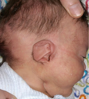
6-Ear anomalies (Figure 3) and/or deafness (Figure 4) 

➧ Diagnosis is made on the presence of at least four of the criteria.

Anesthetic Management of Pt. with CHARGE Syndrome
Figure 1: Coloboma of iris

Anesthetic Management of Pt. with CHARGE Syndrome
Figure 2: Atresia of the choanae

Anesthetic Management of Pt. with CHARGE Syndrome
Figure 3: Ear anomalies

Anesthetic Management of Pt. with CHARGE Syndrome
Figure 4: Deafness (BAHA)

➧ Other abnormalities include:

Muscular hypotonia, facial palsy, tracheo-oesophageal fistula, cleft lip and palate, micrognathia, laryngomalacia, pharyngolaryngeal hypotonia (inability to maintain the patency of the pharyngolaryngeal passage), subglottic stenosis and other upper airway abnormalities. 

➧ There is a high incidence of abnormal blood gas levels and sleep problems. Cardiorespiratory arrest is common in this group of patients. 

➧ Gastroesophageal reflux has been reported. 

➧ Anesthesia may be required for choanal atresia repair, cardiac surgery, tracheoesophageal fistula, ear surgery, Nissen’s fundoplication, and tracheostomy.

Anesthetic Management:

Preoperative Management: 

1. Preoperative assessment of congenital cardiac defects. 

2. Preoperative assessment of upper airway abnormalities. Pharyngo-laryngeal hypotonia causes variable obstruction, which becomes more pronounced during sleep and during inspiration. 

3. Precautions against aspiration of gastric contents. 

Intraoperative Management: 

1. A range of sizes of endotracheal tubes should be available due to subglottic stenosis. 

2. If micrognathia is present, inhalational induction is advisable. 

3. Tendency for upper airway collapse during light anesthesia due to laryngomalacia or pharyngolaryngeal hypotonia. Edematous arytenoids may result from gastroesophageal reflux. 

4. Tracheal intubation difficulties have been recorded and intubation problems are increased with increasing age. 

5. Tracheostomy may be required for long-term management. Some authors felt that early tracheostomy helped to avoid hypoxemic events in infancy. 

Postoperative Management: 

1. Postoperative monitoring of apnea. 

2. Feeding difficulties and a high incidence of gastroesophageal reflux. 

Postoperative Mortality: 

1. Apnea due to pharyngolaryngeal hypotonia. 

2. Postoperative deaths were frequently associated with pulmonary aspiration. 

3. Patients require multiple anesthetics, with an increased incidence of postoperative mortality.

Ultrasound Artifacts

Ultrasound Artifacts


Ultrasound Artifacts


1-Reverberation artifact:

➧ The processing unit in the ultrasound machine assumes echoes return directly to the processor from the point of reflection. 

➧ Depth is calculated as D = V × T, where V is the speed of sound in biological tissue and is assumed to be 1,540 m/sec, and T is time. 

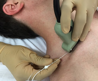
➧ In a reverberation artifact, the ultrasound waves bounce back and forth between two interfaces (the lumen of the needle) before returning to the transducer. 

➧ Since velocity is assumed to be constant at 1,540 m/sec by the processor, the delay in the return of these echoes is interpreted as another structure deep into the needle and hence the multiple hyperechoic lines beneath the block needle (Figure 1). 

Ultrasound Artifacts
Figure 1: Reverberation artifact


2-Mirror artifact:

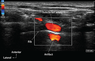
➧ A mirror artifact is a type of reverberation artifact. 

➧ The ultrasound waves bounce back and forth in the lumen of a large vessel (subclavian artery). 

➧ The delay in the time of returning waves to the processor is interpreted by the machine as another vessel distal to the actual vessel (Figure 2). 

Ultrasound Artifacts
Figure 2: Mirror artifact


3-Bayonet artifact:

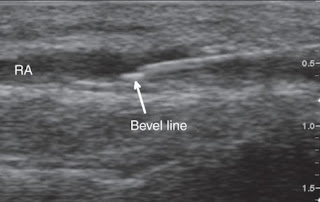
➧ The processor assumes that the ultrasound waves travel at 1,540 m/sec through biological tissue. However, we know that there are slight differences in the speed of ultrasound through different biological tissues. 

➧ The delay in the return of echoes from tissue that has a slower transmission speed, coupled with the processor’s assumption that the speed of ultrasound is constant, causes the processor to interpret these later returning echoes from the tip of the needle traveling in tissue with slower transmission speed as being from a deeper structure and thus giving a bayoneted appearance. 

➧ If the tip is traveling through tissue that has a faster transmission speed, then the bayoneted portion will appear closer to the transducer (Figure 3). 

Ultrasound Artifacts
Figure 3: Bayonet artifact


4-Acoustic Enhancement artifact:

➧ Acoustic enhancement artifacts occur distal to areas where ultrasound waves have traveled through a medium that is a weak attenuator, such as a large blood vessel. 

➧ Enhancement artifacts are typically seen distal to the femoral and the axillary artery (Figure 4). 

Ultrasound Artifacts
Figure 4: Acoustic enhancement artifact


5-Acoustic shadowing:

➧ Tissues with high attenuation coefficients, such as bone, do not allow the passage of ultrasound waves. 

➧ Therefore any structure lying behind tissue with a high attenuation coefficient cannot be imaged and will be seen as an anechoic region. (Figure 5). 

Ultrasound Artifacts
Figure 5: Acoustic shadowing artifact


6-Absent blood flow:

➧The Color-Flow Doppler may not detect blood flow when the ultrasound probe is perpendicular to the direction of blood flow (Figure 6). 

➧ A small tilt of the probe away from the perpendicular should visualize the blood flow (Figure 7, Figure 8). 

➧ Alternatively, for deep vascular structures, signals may be lost due to attenuation. 

➧ Increasing gain, while in Doppler Color-Flow mode, will increase the intensity of the returning signals, which may detect blood flow that was not previously detected. 

Ultrasound Artifacts
Figure 6: Radial a. Absent blood flow artifact
Ultrasound Artifacts
Figure 7: Radial a. Probe tilted away from the direction of blood flow
Ultrasound Artifacts
Figure 8: Radial a. Probe tilted towards the direction of blood flow

Drugs with Rebound Phenomenon

Drugs with Rebound Phenomenon


Drugs with Rebound Phenomenon
➧ The rebound effect, or rebound phenomenon, is the tendency of some medications, in sudden discontinuation, to cause a return of the symptoms it relieved, to a degree stronger than they were before treatment first began. Medications with a known rebound effect can be withdrawn gradually, or, in conjunction with another medication that does not exhibit a rebound effect. 


1-Sedative Hypnotics: 

-Benzodiazepine withdrawal can cause rebound anxiety and insomnia. 

-Eszopiclone and Zolpidem) can cause rebound insomnia. 

2-Stimulants:

➧ e.g. Methylphenidate or Dextroamphetamine 

➧ Rebound effects include psychosis, depression, and a return of ADHD symptoms but in a temporarily exaggerated form. 

3-Antidepressants:

➧ e.g. SSRIs

➧ Cause rebound depression and/or panic attacks and anxiety when discontinued. 

4-Alpha-2 adrenergic agents:

➧ e.g. Clonidine and Guanfacine

➧ The most notable rebound effect is rebound hypertension. 

5-Beta-adrenergic antagonists:

➧ e.g. Bisoprolol

➧ Sudden withdrawal leads to rebound tachycardia and anginal pain. 

6-Highly potent corticosteroids:

➧ e.g. Clobetasol for psoriasis

➧ Abrupt withdrawal can cause rebound psoriasis and hypoglycemia. 

7-Warfarin: 

➧ Withdrawal leads to thromboembolism 

8-Alcohol:

➧ Withdrawal leads to alcohol withdrawal syndrome: (anxiety and convulsions). 

9- Painkillers:

➧ Withdrawal can cause rebound headaches. 

10-Topical decongestants:

➧ Nasal sprays e.g. Phenylephrine

➧ Continuous usage can lead to constant nasal congestion, known as Rhinitis medicamentosa, and discontinuation to rebound nasal congestion.

Drugs affecting IOP

Drugs affecting IOP


Drugs affecting IOP
➧ Normal intraocular pressure (IOP) is between (10 - 20 mmHg). The average value of IOP is 15.5 mmHg with fluctuations of about 2.75 mmHg.

➧ IOP also varies with other factors such as heart rate, respiration, fluid intake, systemic medication, and topical drugs.

➧ Intraocular vascular tone is predominantly affected by CO₂; hypocarbia decreases IOP through vasoconstriction of the choroidal blood vessels and decreases the formation of aqueous humor through reduced carbonic anhydrase activity. The increased IOP associated with hypoventilation and hypercarbia occurs as a result of vasodilation of CBV and increases in central venous pressure. 

A) Drugs that ↑ IOP:

1-Steroid-induced glaucoma:

Mechanism:

Is a form of open-angle glaucoma that is usually associated with topical steroid use, but it may develop with inhaled, oral, intravenous, periocular, or intravitreal steroid administration. 

Risk factors:

-Preexisting primary open-angle glaucoma

-Family history of glaucoma

-High myopia, diabetes mellitus

-History of connective tissue disease (especially rheumatoid arthritis). 

➧ Patients on chronic corticosteroid therapy can remain undiagnosed with an elevated IOP, which can result in glaucomatous optic nerve damage. 

➧ Steroid-induced IOP elevation typically occurs within a few weeks of beginning steroid therapy. In most cases, the IOP lowers spontaneously to the baseline within a few weeks to months upon stopping the steroid. In rare instances, the IOP remains elevated.

2-Topical anticholinergic or sympathomimetic dilating drops, TCA, MAOI, antihistamines, antiparkinsonian drugs, antipsychotic medications, and antispasmolytic agents:

Mechanism:

These medications produce pupillary dilation and precipitate an attack of acute angle-closure glaucoma in anatomically predisposed eyes that have narrow angles. 

3-Sulfa containing medications:

Mechanism:

Induce anterior rotation of the ciliary body causing angle-closure glaucoma. Typically, the angle-closure is bilateral and occurs within the first several doses of the sulfonamide-containing medication. Patients with narrow or wide-open angles are potentially susceptible to this rare and idiosyncratic reaction.

4-Ketamine: 

The effect on IOP varies. Early studies reported an increase in IOP after IV or IM administration of ketamine. Ketamine given after premedication with diazepam and meperidine does not affect IOP and IM administered ketamine may even lower IOP in children. 

5-Depolarizing muscle relaxants (Succinylcholine):

Causes a transient (4–6 min) but significant increase in IOP of (10 - 20 mm Hg). Although the mechanism is unclear, the increase is not attributable simply to induced muscle fasciculations. 

6-Large volume Local anesthetic:

Injecting a large volume (8–10 mL) of Local anesthetic into the orbit (e.g. peribulbar block).

7-Tracheal intubation:

Sympathetic cardiovascular responses to tracheal intubation. 

8- Caffeine

B) Drugs that ↓ IOP:

In general, CNS depressants lower IOP.

1-Intravenous anesthetics and volatile agents:

Mechanism:

Relax extraocular muscle tone, depress the CNS (i.e., the diencephalon), improve the outflow of aqueous humor, and lower venous and arterial blood pressures.

➧ e.g. thiopental, propofol, etomidate, decrease in IOP by 14 - 50 % have been noted.

➧ During controlled ventilation and normocapnia, volatile inhaled anesthetics reduce IOP in proportion to the depth of anesthesia.

2-Non depolarizing neuromuscular blocking drugs:

Either do not affect IOP or produce a slight decrease.

3-Benzodiazepines:

IV administered diazepam (0.15 mg/kg) and equipotent intravenous doses of midazolam (0.03 mg/kg).

4-Narcotic premedication:

Causes no change, or only a slight decrease, in IOP. 

5-Neuroleptanalgesia:

Produced by mixtures of (fentanyl and droperidol) decreases IOP by 12 % in normocapnic patients. 

6- Alcohol consumption:

This leads to a transient decrease in IOP.

7- Several pretreatment regimens:

➧ IV lidocaine (1.5 mg/kg) or sufentanil (0.05–0.15 ”g/kg) given 3 - 5 min. before induction. 

➧ Oral administration of the centrally acting antihypertensive drug clonidine (5 ”g/kg) 2 hrs before induction of anesthesia blunts the IOP response to intubation. 

➧ Intranasal administration of nitroglycerin

➧ ÎČ-adrenergic receptor blocking drugs

Drugs Avoided in Patients with Renal Failure

Drugs Avoided in Patients with Renal Failure


Drugs Avoided in Patients with Renal Failure


➧ The excretion of water-soluble drugs and their active metabolites will be impaired. For drugs that are renally excreted the half-life increases slowly with deteriorating renal function until severe nephron loss at which point the half-life increases sharply with further reductions in renal function. Dialysis can only usually replace a small part of the excretory capacity of the healthy kidney. 

Antibacterial agents:

1-Injectable penicillin G or carbenicillin: may be associated with neuromuscular toxicity, myoclonus, seizures, or coma. 

2-Vancomycin 

3-Amphotericin 

4-Tetracyclines: except doxycycline (Vibramycin), have an antianabolic effect that may significantly worsen the uremic state in patients with severe disease. 

5-Aminoglycosides 

6-Imipenem/cilastatin (Primaxin): can accumulate in patients with chronic kidney disease, causing seizures if doses are not reduced. 

7-Sulphonamides 

8-Nitrofurantoin (Furadantin): has a toxic metabolite that can accumulate in patients with chronic kidney disease, causing peripheral neuritis. 

Anesthetic drugs:

1-Muscle relaxants:

Depolarizing muscle relaxant:

-Suxamethonium: should be avoided if hyperkalemia is present. 

Non-depolarising muscle relaxants (NDMRs): 

-NDMRs depends on the kidney for elimination)

-Gallamine: should be avoided 

-Pancuronium, pipecuronium, alcuronium, curare, and doxacurium: should be used with caution. Potentiation of neuromuscular blockade may occur in the presence of metabolic acidosis, hypokalemia, hypermagnesemia, or hypocalcemia and with medications such as aminoglycosides. Monitor neuromuscular blockade whenever possible. 

-Vecuronium and mivacurium: are safe to use in renal failure as only small percentages are excreted renally.

2-Opioids:

-Morphine: is metabolized in the liver to morphine-6-glucuronide which has about half the sedative effect of morphine with a markedly prolonged half-life. 

-Pethidine: is partially metabolized to norpethidine which is less analgesic and has excitatory and convulsant properties. 

-Tramadol and codeine: Metabolites can accumulate in patients with chronic kidney disease, causing central nervous system and respiratory adverse effects.

3-Inhalational agents:

➧ There is decreased elimination of the fluoride ions which are significant metabolites of enflurane, sevoflurane, and methoxyflurane which can worsen renal function, so these inhalational agents should be avoided especially if used at low flows. 

Non-Steroidal Anti-Inflammatory Drugs (NSAIDs):

➧ NSAIDs should be avoided as all decrease renal blood flow and may precipitate complete renal failure. 

➧ Adverse renal effects of NSAIDs include acute renal failure; nephrotic syndrome with interstitial nephritis; and chronic renal failure.

➧ The risk of acute renal failure is three times higher in NSAID users than in non-NSAID users.

➧ Other adverse effects of NSAIDs include decreased potassium excretion, which can cause hyperkalemia, and decreased sodium excretion, which can cause peripheral edema, elevated blood pressure, and decompensation of heart failure.

➧ NSAIDs can blunt antihypertensive treatment, especially if beta-blockers, ACE inhibitors, or ARBs are used.

Management of Failed Spinal Anesthesia

Management of Failed Spinal Anesthesia


Management of Failed Spinal Anesthesia
➧ Failure of a spinal anesthetic is an event of significant concern for both patient and anesthetist when it is immediately apparent, but it can have serious consequences (clinical and medico-legal) if the problem only becomes evident once surgery has started.

➧ This can be a source of pain, anxiety, and psychological trauma to the patient and a cause of stress, complaints, and medico-legal sequelae to the anesthetist.

Prevention is better than cure:

➧ The spinal block should be performed with meticulous attention to detail. 

➧ If there is any doubt about the nature or duration of the proposed surgery, a method other than standard spinal anesthesia should be used.

Management of failed spinal block:

-The precise management of failed spinal block will depend on the nature of the inadequacy and the time at which it becomes apparent. 

-The slower the onset of either motor or sensory block, the more likely is the block to be inadequate, so the more detailed assessment should be. 

-While the onset of spinal anesthesia is rapid in most patients, it can be slow in some; so, ‘transient time’ should always be allowed. 

-However, if the expected block has not developed within 15 min., some additional maneuver is needed, as follows:

1-No block:

Causes:

-Incorrect or ineffective solution was injected. 

-Solution has been deposited in the wrong place. 

Management: 

-Repeating the block or conversion to general anesthesia is the only option.

2-Spinal block of inadequate height:

Causes:

-Some injectate has been lost or misplaced. 

-The level of injection was too low.

-Anatomical abnormality has restricted spread.

Management:

-If a hyperbaric solution was used, flex the patient’s hips and knees and tilt the table head down (Trendelenburg position). This straightens out the lumbar lordosis, but maintains a cephalad ‘slope’ and allows any solution ‘trapped’ in the sacrum to spread further. 

-In an obstetric situation, turn the patient to the full lateral position with a head-down tilt and reverse the side after 2–3 min.

-If a plain (and usually slightly hypobaric) solution has been used, it may help to sit the patient up but beware of peripheral pooling of blood.

-If an intrathecal catheter injection results in an inadequate spread, do not inject more of the same solution because the dose has minimal effect on the intrathecal spread. 

-Either posture should be manipulated as above, or a different baricity of solution should be tried, or the catheter should be withdrawn before the injection is repeated.

3-Unilateral block:

Causes: 

-This is most likely because of positioning. 

-The longitudinal ligaments supporting the cord have blocked spread. 

Management:

-If the operation is on the anesthetized limb, the surgeon should know that the other leg has sensation, and the patient should be reassured and closely monitored. 

-Otherwise, turning the patient onto the unblocked side if a hyperbaric solution was used (or the reverse for plain solutions) may facilitate spread.

4-Patchy block:

The block appears adequate in extent, but the sensory and motor effects are incomplete. 

Causes: 

-The local anesthetic (LA) dose was inadequate.

-The LA was partially misplaced.

Management:

-If this becomes apparent before surgery starts, the options are to repeat the spinal injection or to use IV analgesia, the latter being the only option after skin incision.

-It may not be necessary to recourse to general anesthesia, as sedation or analgesic drugs are often sufficient especially when patient anxiety is a major factor. 

-Infiltration of the wound and other tissues with LA by the surgeon may also be useful in such situations.

5-Inadequate duration:

Causes:

-An inadequate dose of LA was delivered to the CSF. 

-Syringe swap; Lidocaine (intended for skin infiltration) was confused for bupivacaine. 

-The operation has taken longer than expected.

Management: 

-Sedation, IV analgesia, or infiltration of LA may be adequate, but often the only option is to convert to general anesthesia.

Repeating the spinal block:

➧ If no effect at all was seen 15-20 min. following the injection, it seems reasonable to repeat the block, paying close attention to avoiding the potential pitfalls. 

➧ In all other situations besides total failure, there must be some LA already in the CSF, and anxieties relating to several issues have to be taken into account: 

1-A restricted block may be due to an anatomical factor, impeding the physical spread of the solution, and it may have the same impact on a second injection, resulting in a high concentration of LA at or close to the site of injection leading to neurotoxicity.

2-Barriers to spread within the subarachnoid space may also affect epidural spread (and vice versa), so an attempt at epidural block may not succeed either.

3-Repeated injection in response to a poor quality block may lead to excessive cephalad spread with the potential for cardiovascular instability, respiratory embarrassment, or total spinal anesthesia, so a lower dose should be used to reduce this risk. 

4-A good quality, but unilateral block, might lead to an attempt to place a second injection into the ‘other’ side of the theca, but the risk of placing the second dose on the same side must be significant. 

5-A block of inadequate cephalad spread might be overcome by repeating the injection at a higher level, but should only be attempted when there is a considerable indication for a regional technique.

6-When a repeat block is considered, the adjacent nerve tissue is already affected by LA action, so the risk of direct needle trauma is increased.

Recourse to general anesthesia:

➧ There are many ways in which an inadequate block might be ‘rescued’: 

-General anesthesia must be considered if one or two simple measures have not rectified matters. 

-Common sense and clinical experience are usually the best indicators of exactly when to convert to general anesthesia. 

-If general anesthesia is induced to supplement a partially effective spinal anesthesia, any degree of sympathetic nerve block will make hypotension more likely.

Postoperative Management:

1-Documentation and follow-up:

-The anesthetic complication details should be fully documented in the notes. 

-The patient should be provided with an apology and a full explanation after the operation. 

-Giving the patient a written summary of events for presentation to a future anesthetist can be very helpful, although care should be taken to prevent medico-legal recourse.

-Rarely, the inadequate spread has been the first indication of pathology within the vertebral canal, so if there is any suspicion, look for symptoms and signs of neurological disease, and consult a neurologist. 

-During follow-up of a patient in whom no block was obtained, the possibility of LA ‘resistance’ may seem an attractive explanation.

2-Investigating LA effectiveness:

-Performing skin infiltration with some of the solutions intended for the spinal injection should demonstrate that it is effective. 

-If the concern continues the operating theater, pharmacy, and anesthetic department records should be cross-checked to see whether other practitioners in the hospital have experienced any problems. 

-Similarly, distributors should be able to check whether other hospitals, which have been supplied with material from the same batch, have reported difficulty.


Failed Spinal Anesthesia

Failed Spinal Anesthesia

Introduction:

➧ Spinal (intrathecal) anesthesia is one of the most reliable regional block methods: the needle insertion technique is relatively straightforward, with cerebrospinal fluid (CSF) providing both a clear endpoint of successful needle placement and a medium for carriage of local anesthetic (LA) within subarachnoid space. However, the possibility of failure has long been recognized, with an incidence of less than 1% in experienced hands.

➧ Literally, the word failure implies that spinal anesthesia was attempted, but no block resulted or a block results, but is inadequate for the proposed surgery. 

➧ Such inadequate block may be related to the three components of the block: the extent, the quality, or the duration of local anesthetic action, often with more than one of these being inadequate.

➧ The intrathecal injection can go astray within each of the five phases of an individual spinal anesthetic, leading to blocking failure, these being, in sequence: 

1-Lumbar puncture 

2-Solution injection 

3-Spreading of the drug through CSF 

4-Drug action on the spinal nerve roots and cord 

5-Subsequent patient management

Causes and prevention of failure:

1-Unsuccessful Lumbar Puncture (Dry Tap):

-Inability to obtain CSF ‘Dry Tap’, is the only cause of failure which is immediately obvious. 

Causes:

a) Incorrect needle insertion or Poor patient positioning:

Prevention: 

➧ A calm, relaxed patient is more likely to assume and maintain the correct position, so: 

-Explanation (before and during the procedure) and gentle, unhurried patient handling are vital.

-Premedication with light anxiolytic for relaxing the patient. 

-Local anesthetic infiltration at the puncture site is effective without obscuring the landmarks.

-Systemic analgesia (IV or inhalation) helps in achieving the correct position for patients in pain (e.g. from a fractured hip).

b) Anatomical abnormalities of the spine:

-Kyphosis, scoliosis, calcification of ligaments, consequences of osteoporosis, obesity, and patient anxiety, make both positioning the patient and needle insertion more difficult, especially in the elderly. 

Prevention: 

-Good clinical training is the key to success.

-Adherence to the basic rules of positioning, needle insertion, and use of adjuncts.

-Lateral or paramedian approach, especially if the mid-line ligaments are heavily calcified. 

-Ultrasound guidance: a pre-procedure scan can be useful in patients with anatomical abnormality to identify the midline and level of injection and to assess the depth of dura from the skin.

c) Equipment-related factors: A blocked needle lumen:

Prevention: 

-Both needle and stylet must be checked for correct fitness before use.

-The needle should not be advanced without the stylet in place, because tissue or blood clots can easily obstruct the fine bore needles used now.

-Prompt needle withdrawal and 'flush test' to assure patency.

2-Pseudo-successful Lumbar Puncture:

-The appearance of clear fluid at the needle hub is usually the final confirmation that the subarachnoid space has been entered.

a) Epidural 'Top-up' dose:

-Rarely, the clear fluid is not CSF, but LA injected as an epidural ‘top-up’ dose or spreading from the lumbar plexus.

Prevention:

-Unfortunately, a positive test for glucose in the fluid does not confirm that this fluid is definitely CSF because extracellular fluid constituents diffuse rapidly into fluids injected into the epidural space.

b) Congenital arachnoid cyst:

-Another, rarer cause is a congenital arachnoid cyst (Tarlov cyst), which are meningeal dilatations of the posterior spinal nerve root, present in 4.5-9% of the population.

3-Solution Injection Errors:

1. Dose selection:

-The dose injected, within the normal range, has only a small effect on the height of a spinal block but is important in determining the quality and duration of the block. 

➧ The dose chosen will depend on: 

-The specific LA used

-The baricity of LA solution

-The patient’s subsequent posture

-The type of block intended

-The extent and duration of planned surgery

Causes: 

➧ Some anesthetists use lower doses than is traditional, in attempts to either: 

-Minimize hypotension, by producing a unilateral block. 

-Decrease block duration which speeds postoperative mobilization and decreases the need for bladder catheterization.

➧ Such lower doses will increase the margin for error and exaggerate the consequences of other problems such as: 

-Loss of injectate and so risk an inadequate block.

-The ‘dead space’ of the needle and hub will contain a significant proportion of what is a small volume to start with.

2. Loss of injectate:

Causes: 

-Leakage of LA solution through the Luer connection between syringe and needle. 

-Leakage through a defect at the junction of needle hub and shaft. 

➧ Given the small volumes involved, the loss of a few drops can cause a significant decrease in the mass of the drug reaching the CSF, and thus in its effectiveness. 

Prevention: 

-Insert the syringe containing the injectate firmly into the hub of the needle, and check that no leakage occurs.

3. Misplaced injection:

Causes: 

➧ Anterior or posterior displacement of the needle tip from subarachnoid to epidural space:

a) During connection of the syringe to the needle, where deposition of a spinal dose of LA will have little or no effect. 

b) During fluid aspiration for confirmation that the needle tip is still in the correct space, may displace the tip unless performed carefully, as may the force of the injection of the syringe contents. 

Prevention:

-The dorsum of one hand should be anchored firmly against the patient’s back and the fingers are used to immobilize the needle, while the other hand is used to manipulate the syringe.

-After attachment of the syringe, aspirate 0.5-1 ml to confirm the free flow of CSF, and at the end of spinal injection, aspirate 0.5-1 ml, to confirm that the needle tip is still in the subarachnoid space, the aspirated volume is re-injected before the needle is withdrawn. Some anesthetists advocate that this is done halfway through as well, 

c) Tip displacement is an important issue with the ‘Pencil point’ needles, as the opening at the end of these needles is proximal to the tip, so only a minor degree of ‘backward’ movement during syringe attachment may result in epidural injection. 

-Also, the opening of these needles may ‘straddle’ the dura so that some solution reaches the CSF, and some of the epidural space. 

-This may be exaggerated by the dura acting as a ‘flap’ valve across the needle opening. Initially, CSF pressure pushes the dura outwards so that aspiration is successful, but subsequent injection pushes the dura forward and the solution is misplaced. 

-A variant is that the needle tip penetrates the dura, but it is the arachnoid mater that acts as the flap valve so that accidental subdural injection results. 

Prevention: 

-Rotation of the needle through 360 degrees after the initial appearance of CSF, and before check aspiration, as the rotation reduces the risk of the membrane edges catching on the opening.

4. Inadequate intrathecal spread:

➧ Factors affecting the intrathecal spread of a local anesthetic solution: 

-Anatomy of the vertebral canal.

-Solution physical characteristics.

-Gravity.

a) Anatomical abnormality: 

-Abnormalities of the curves of the vertebral column as kyphosis or scoliosis, may interfere with the solution spread.

Examination of the patient should reveal whether this might occur, but it is not possible to predict whether the effect will be excessive spread or failure. 

-A rare possibility, is that the ligaments supporting the spinal cord within the theca, form complete septae which act as longitudinal or transverse barriers to LA spread. This can result in a block that is entirely unilateral or limited cephalad spread.

-Spinal stenosis or other pathological lesions can limit spread, effectiveness, or both.

-Previous spinal surgery or intrathecal chemotherapy may result in adhesions that interfere with LA spread. 

-Increased CSF volume in the lumbar theca can cause restricted cephalad spread of intrathecal injection.

-A variation of this factor is dural ectasia, which is a pathological enlargement of the dura seen in most patients with Marfan’s syndrome and in some other connective tissue disorders. 

b) Solution density (baricity):

-Isobaric solutions, with a density within the normal range of CSF, will block the lower limbs with little risk of thoracic nerve block and thus less hypotension. 

-Plain solutions of bupivacaine, although referred to as isobaric, are actually of lower density to be hypobaric at body temperature (37ÂșC). They have a less predictable spread than that of a truly isobaric preparation, and the block may be not higher than the second lumbar dermatome with slow onset. 

-Hyperbaric solutions, with a density greater than that of CSF, move under the combined influence of gravity and the curves of the vertebral canal. If the patient is placed supine after the injection of a hyperbaric preparation at the mid-lumbar level, the solution will spread ‘down’ the slope under the effect of gravity to pool at the ‘lowest’ point of the thoracic curve, so exposing all nerve roots up to that level to an effective concentration of LA. (Figure 1)

-However, if a lumbar puncture is performed at the fourth lumbar or the lumbosacral interspace, LA may be ‘trapped’ below the lumbar curve, especially if the patient is in the sitting position during injection and maintained in that position for a period thereafter. This results in a block that is restricted to the sacral segments.

Effects of drug baricity
Figure 1: Effects of drug baricity
Prevention:

-Avoiding too low injection level unless a deliberate ‘saddle’ block is intended.

5. Ineffective drug action:

➧ The solution injected reaches the target nerves, but it is ineffective or inactive, with a variety of possibilities: 

a) Incorrect drug injection (Identification error): 

-Using LA for skin infiltration or analgesic adjuvants, used from the same sterile preparation area, instead of spinal LA, may lead to an ineffective block. 

Prevention:

-The use of labeled syringes, but this is not easy within a sterile field. 

-The use of syringes with different sizes for each component of the procedure.

-Minimizing the number of ampoules on the block tray (such as using the same LA for both skin infiltration and spinal anesthesia).

b) Physico-chemical incompatibility: 

-The mixing of two different pharmaceutical preparations raises the possibility of ineffectiveness as a result of interaction between LA and adjuvant.

-Chemical reaction can generate an obvious precipitate, or lower the pH of the LA solution which will decrease the concentration of the unionized fraction which diffuses into nerve tissue resulting in a decreased effect.

-Local anesthetics are compatible with most opioids, but the situation is less definitive with other adjuvants such as clonidine, midazolam, ketamine, and other substances. 

-The stability is unknown when mixing three or more substances together for intrathecal use. 

-The incidence of failure is greater after the addition of a vasoconstrictor solution. 

c) Inactive LA solution: 

-Ester-type LAs, are chemically labile so that heat sterilization and prolonged storage, particularly an aqueous solution, can make them ineffective because of hydrolysis, and hence they need very careful handling. 

-Amide-type LAs (e.g. lidocaine, bupivacaine, etc.) are more stable and can be heat sterilized and stored for several years without loss of potency. 

d) Local anesthetic resistance: 

-Very rarely, failed spinal block has been attributed to physiological resistance to the actions of LA drugs.

-This problem is due to mutation of sodium channels (channelopathy) which is associated with significant neurological diseases such as; intractable epilepsy and chronic pain, however, this does not exist in asymptomatic individuals.

-A history of repeated failure of dental or other LA techniques is accompanied by speculation that the problem is due to sodium channel mutation that renders the drugs ineffective. 
4-Failure of Subsequent management:

➧ Not all of a patient’s claims of discomfort, or pain, during spinal anesthesia, are due to inadequate block.

Causes:

-Lying awake during surgery is not a pleasant experience for most patients, and anxiety alone can cause patient discomfort. 

-Furthermore, operating tables are designed for surgical access, not patient comfort; and intra-abdominal stimuli can result in afferent impulses in unblocked parasympathetic nerve fibers causing unpleasant sensations. 

Prevention: 

-Good preoperative patient counseling followed by a supportive approach from the anesthetist during the operation is important in avoiding such problems. 

-Judicious, and proactive use of systemic analgesic drugs. 

-Sufficient sedation to produce drowsiness, or even sleep (with appropriate monitoring), is indicated except in obstetric situations, where small doses may be useful.

-Distraction techniques such as listening to music.

5-Testing the block:

-It is mandatory to test the level of the block before surgery commences

-Most patients will have some anxiety about the effectiveness of the injection, and this will be increased if testing is started too soon. 

-Conventional practice is to check motor block by testing the ability to lift the legs, followed by testing of sensory block to stimuli such as light touch, cold, or pin-prick.

-It is advisable to start testing in the lower segments, where onset will be fastest, and work upwards. Proving early that there is some effect encourages patient confidence; testing too soon does the opposite. 

-Establishing that the level of block is appropriate for the projected surgery is often taken to demonstrate that the quality of block is adequate also. 

-A covert pinch of the site of the proposed surgical incision may be a better indicator of skin analgesia and can be reassuring if the block has been slow in onset. Asking the surgeon to do the same with toothed surgical forceps after distracting the patient with conversation.

6-Catheter and Combined Techniques:

-The majority of spinal anesthetics involve a single shot of LAs. 

-To take advantage of the rapid onset and profound block of spinal anesthesia, both continuous and combined spinal-epidural techniques have been introduced to increase flexibility. 

-If the catheters are correctly placed, problems of inadequate spread, quality, and duration of effect can be dealt with. 

-However, insertion of an intrathecal catheter can be difficult to achieve in some patients and can result in the misdirection of the LA solution, with the risk of neurotoxicity.


Read more: ☛ Failed Epidural Block