Accidental Subdural Injection of Local Anesthetics
Predisposing factors:
1-Difficult epidural block.
2-Previous back surgery.
3-Recent lumbar puncture (CSF leak through epidural rent leading to distended subdural space).
4-Rotation of the epidural needle in epidural space through an arc of 180°.
5-Prolonged epidural catheterization.
Characterized by:
1-Subdural space may not be detected by aspiration test or test dose.
2-Delayed onset, Short duration.
3-Extensive block spread (Block is disproportionate to the amount of LA injected).
4-Segmental, Asymmetric, Patchy distribution.
5-Cranial spread rather than caudal spread.
6-High sensory block can involve cranial nerves.
7-Sparing of sacral roots.
8-Minimal motor blockade.
9-Relative lack of sympathetic block.
N.B. Subdural space has more potential capacity posterior & lateral. There is rarely motor paralysis or severe hypotension due to the sparing of anterior nerve roots that transmit motor & sympathetic fibers.
Detection of subdural placement of epidural catheter:
1-Stimulation test (using nerve stimulator):
 |
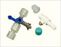
| Figure 1: Johns ECG adaptor |
➧ A nerve stimulator is connected to the epidural catheter via an adapter (Johans ECG adapter, Arrow International, Inc., Reading, USA), (Figure 1).
➧ The epidural catheter and ECG adapter are primed with sterile normal saline.
➧ The anode lead of the nerve stimulator is connected to an electrode over the upper or lower extremity as a grounding site.
➧ The cathode lead of the stimulator is connected to the metal hub of the adapter.
➧ The nerve stimulator is set at a frequency of 1 Hz with a pulse width of 200 msec.
➧ Electrical stimulation (1-10 mA) with a segmental motor response (truncal or extremities movement) indicates that the catheter is in the epidural space.
➧ No motor response indicates that it is not.
➧ Since it is possible to obtain vigorous and uncomfortable twitches with an excessive current, the current output must be carefully increased from zero and stopped once the motor activity is visible.
➧ Thus, the stimulator used in the test must be sensitive enough to allow a gradual increase of current output from zero up to at least 10 mA.
➧ Because a motor response will be elicited at a very low current (< 1 mA) in the case of subarachnoid or subdural placement, the current output must be carefully increased in small increments (0.1 mA) between zero and 2 mA.
2-Contrast Fluoroscopy:
➧ Injection of 3 ml contrast medium via the epidural catheter then radiography of spinal cord is done.
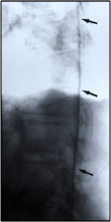
➧ If the epidural catheter is in subdural space, the lateral view will show a contrast medium spreading cephalad over many segments, along the dorsal part of the spinal cord (Figure 2).
 |
| Figure 2: Contrast Fluoroscopy, Lateral view |


