Educational Blog about Anesthesia, Intensive care and Pain management

Showing posts with label Pediatric anesthesia. Show all posts

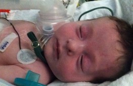
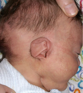
Treacher Collins Syndrome

Treacher Collins Syndrome:


Treacher Collins Syndrome
-A craniofacial defect associated with developmental anomalies of the first arch. 

-Abnormalities vary from minimal, to complete syndrome. 

-The syndrome is named after Edward Treacher Collins, an English surgeon and ophthalmologist, who described its essential traits in 1900. 

-Patients may require anesthesia for maneuvers to improve upper airway obstruction temporarily, or for correction of some of the congenital defects. 

-Airway obstruction and a requirement for multiple operations increase the need for tracheostomy. 

Etiology:

-It is due to mutations in TCOF1, POLR1C, or POLR1D genes. TCOF1 gene mutations are the most common cause of the disorder, accounting for 81 to 93% of all cases. POLR1C and POLR1D gene mutations cause an additional 2% of cases. In individuals without an identified mutation in one of these genes, the genetic cause of the condition is unknown. 

Preoperative Abnormalities:

1. Features may include mandibular and malar hypoplasia, antimongoloid palpebral fissure, macrostomia, irregular maloccluded teeth, microphthalmia, lower lid defects, cleft palate, high arched palate, macroglossia, and auricular deformities. 

2. Associated abnormalities include mental retardation, deafness, dwarfism, cardiac defects, choanal atresia, and skeletal deformities. 

3. Chronic upper respiratory tract obstruction and obstructive sleep apnea, which can lead to growth retardation and/or cor-pulmonale. 

Anesthetic Problems:

1. Upper airway obstruction: in neonates, this may require urgent temporary maneuvers, such as stitching the tongue to the lower lip 

2. Excess secretions: may impede induction of anesthesia 

3. Inhalational induction: may be difficult 

4. Difficult tracheal intubation 

5. Obstructive sleep apnea may occur postoperatively 

6. Pulmonary edema 

Intraoperative Management:

Recommendations:

1-Monitoring by pulse oximetry, to detect airway obstruction, is crucial. 

2-Avoid respiratory depressant drugs, both for premedication and postoperatively 

3-Use drying agents 

4-Never give muscle relaxant until the airway has been secured. 

Airway Management:

-Several methods have been proposed to overcome the problem of difficult intubation, some in the awake patient and some under general anesthesia: 

a) In Awake Patients: 

1-Awake intubation or awake direct laryngoscopy to visualize the vocal cords. 

2-Direct laryngoscopy, with the patient in the sitting position, and using a 5-G feeding tube taped to the side of the laryngoscope to give oxygen. 

3-The Augustine guide (Figure 1): can be used for nasotracheal intubation in an awake, sedated patient.
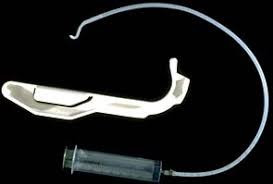
Augustine guide
Figure 1: Augustine guide

4-Fibreoptic bronchoscope or Tracheostomy under local anesthesia: 

In small infants, the ‘tube over bronchoscope’ technique is not always possible because of the small size of the tube, therefore a Seldinger type approach may be necessary: 

-After the administration of atropine, ketamine IM, and topical lidocaine, a fibreoptic bronchoscope (OD 3.6mm, L 60 cm, and suction channel 1.2 mm) was passed through one nostril. 

-The tongue was held forward with Magill forceps, until the vocal cords were seen, but not entered, because of the risk of total obstruction. 

-Under direct vision, a Teflon-coated guidewire with a flexible tip was passed via the suction channel into the trachea. 

-The bronchoscope was carefully removed leaving the wire in place, and an ID 3mm nasotracheal tube was then passed over it into the trachea. 

Pediatric bronchoscopes of 2.5 mm diameter are now available, but their very fineness makes them less easy to handle than the 4-mm bronchoscopes). 

b) In Anesthetized Patients: 

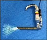
1-Jackson anterior commissure laryngoscope (Figure 2): 

-The head is elevated above the shoulders, with flexion of the lower cervical vertebrae and extension at the atlanto-occipital joint. The laryngoscope is introduced into the right side of the mouth. Only the tip is directed towards the midline, the proximal end remaining laterally so that a further 30 degrees of anterior angulation can be obtained. 

-The narrow, closed blade prevents the tongue from falling in and obscuring the view of the larynx. When visualized, the epiglottis is elevated, and the larynx entered. Intubation is then achieved by passing a lubricated tube, without its adaptor, down the laryngoscope. It is held in place with alligator forceps whilst the laryngoscope is withdrawn.
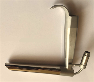
Jackson anterior commissure laryngoscope
Figure 2: Jackson anterior commissure laryngoscope

2-The Bullard intubating laryngoscope (Figure 3):

-It can be used to achieve nasotracheal intubation.
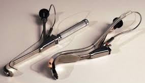
Bullard intubating laryngoscope
Figure 3: Bullard intubating laryngoscope

3-Tactile nasal intubation: 

-Inhalational induction with sevoflurane, and the tongue is pulled downwards and forwards. The tube is initially used as a nasal airway, whilst the index and middle finger are used to palpate the epiglottis, through which the tube is then passed. 

4-Laryngeal mask airway: 

-Inserted under propofol anesthesia and can be used as a conduit for the passage of a fibreoptic bronchoscope.

5-The use of an assistant to pull out the tongue with Magill forceps, and at the same time to apply cricoid pressure, to assist laryngoscopy. 

Postoperative Management:

-The tracheal tube should remain in place until the patient is fully awake. 

-Patients should be nursed in a high dependency area postoperatively. The combination of sleep apnea and drugs with CNS-depressant effects may make them particularly susceptible to respiratory arrest.

Read more ☛ Pierre Robin Syndrome

CHARGE Syndrome

Anesthetic Management of Pt. with CHARGE Syndrome
Definition:

➧ A syndrome characterized by: 

1-Coloboma of the eye (Figure 1) 

2-Heart defects (ASD, VSD, PDA, TOF, Rt. Aortic arch, Double outlet Rt. ventricle) 

3-Atresia of the choanae (Figure 2) 

4-Retarded growth development and/or central nervous system abnormalities 

Severe sensorineural, visual, and vestibular deficits are suggested as the cause of delay in walking development, rather than retardation. 

5-Genital hypoplasia in males (Hypogonadism) 

6-Ear anomalies (Figure 3) and/or deafness (Figure 4) 

➧ Diagnosis is made on the presence of at least four of the criteria.

Anesthetic Management of Pt. with CHARGE Syndrome
Figure 1: Coloboma of iris

Anesthetic Management of Pt. with CHARGE Syndrome
Figure 2: Atresia of the choanae

Anesthetic Management of Pt. with CHARGE Syndrome
Figure 3: Ear anomalies

Anesthetic Management of Pt. with CHARGE Syndrome
Figure 4: Deafness (BAHA)

➧ Other abnormalities include:

Muscular hypotonia, facial palsy, tracheo-oesophageal fistula, cleft lip and palate, micrognathia, laryngomalacia, pharyngolaryngeal hypotonia (inability to maintain the patency of the pharyngolaryngeal passage), subglottic stenosis and other upper airway abnormalities. 

➧ There is a high incidence of abnormal blood gas levels and sleep problems. Cardiorespiratory arrest is common in this group of patients. 

➧ Gastroesophageal reflux has been reported. 

➧ Anesthesia may be required for choanal atresia repair, cardiac surgery, tracheoesophageal fistula, ear surgery, Nissen’s fundoplication, and tracheostomy.

Anesthetic Management:

Preoperative Management: 

1. Preoperative assessment of congenital cardiac defects. 

2. Preoperative assessment of upper airway abnormalities. Pharyngo-laryngeal hypotonia causes variable obstruction, which becomes more pronounced during sleep and during inspiration. 

3. Precautions against aspiration of gastric contents. 

Intraoperative Management: 

1. A range of sizes of endotracheal tubes should be available due to subglottic stenosis. 

2. If micrognathia is present, inhalational induction is advisable. 

3. Tendency for upper airway collapse during light anesthesia due to laryngomalacia or pharyngolaryngeal hypotonia. Edematous arytenoids may result from gastroesophageal reflux. 

4. Tracheal intubation difficulties have been recorded and intubation problems are increased with increasing age. 

5. Tracheostomy may be required for long-term management. Some authors felt that early tracheostomy helped to avoid hypoxemic events in infancy. 

Postoperative Management: 

1. Postoperative monitoring of apnea. 

2. Feeding difficulties and a high incidence of gastroesophageal reflux. 

Postoperative Mortality: 

1. Apnea due to pharyngolaryngeal hypotonia. 

2. Postoperative deaths were frequently associated with pulmonary aspiration. 

3. Patients require multiple anesthetics, with an increased incidence of postoperative mortality.

Pierre Robin syndrome

Pierre Robin syndrome:

Pierre Robin syndrome

Manifestations:

➧ A rare syndrome in which there is a combination of:

-Severe micrognathia

-Posterior prolapse of the tongue


Other congenital abnormalities:

-Cleft palate

-Esophageal atresia

➧ The term syndrome is now reserved for those errors of morphogenesis with the simultaneous presence of multiple anomalies caused by a single etiology.

➧ The term sequence has been introduced to include any condition that includes a series of anomalies caused by a cascade of events initiated by a single malformation.

Pierre Robin sequence:

➧ The common features of which include:

1-Mandibular hypoplasia

2-Glossoptosis (Figure 1)

3-Incomplete cleft palate (Figure 2)

Although all three are not necessarily present.

This results in:

-Respiratory obstruction in infancy

-Failure to thrive

-Occasionally cor-pulmonal


Glossoptosis
Figure 1: Glossoptosis


Incomplete cleft palate
Figure 2: Incomplete cleft palate


➧ The Robin sequence may be an isolated abnormality, or it may be part of a syndrome. There may be airway and intubation problems in any of these patients.

Associated syndromes:

➧ Pierre Robin sequence has been reported as occurring in association with:

-Stickler syndrome (20%-25% of these cases)

-Campomelic dysplasia

-Trisomy 11q syndrome

-Deletion 4q syndrome


-Velocardiofacial syndrome

-Treacher-Collins syndrome.

➧ These patients frequently require anesthesia at a young age.

➧ Management of long-term airway obstruction is a matter of debate, but at present tracheostomy seems to be back in favor. The mortality from pediatric tracheostomy has declined and it increases the safety of subsequent anesthetics.

Etiology:

➧ The exact causes of the Pierre Robin sequence are unknown. Possible mechanisms include:

-Genetic causes.

-Oligohydramnios, which may limit chin growth.

-Weakness of the facial muscles (myotonia).

-Connective tissue disease.

➧ The genetic causes for some of the isolated cases (Pierre Robin sequence without any associated malformations) may include mutations or deletions of parts of the DNA neighboring the SOX9 gene (located in chromosome 17 (17q24)). This gene provides instructions for making protein SOX9 that regulates the activity of other genes, especially those involved in the development of the skeleton, including the jaw during embryonic development.

Preoperative abnormalities:

1. Many present as difficult or failed intubation during resuscitation at delivery. Hypoxic brain damage may be sustained at this stage.

2. The remainder usually presents within a few hours of birth when the micrognathia and glossoptosis cause breathing and feeding difficulties, with episodes of cyanosis when the child is in the supine position. Feeding difficulties correlate with the severity of airway obstruction. Subsequently, there is a failure to thrive.

3. There was a high incidence of concomitant problems, which included gastroesophageal reflux, congenital heart disease, and pulmonary disease.

4. Jaw index: A new index for defining micrognathia by measurement of three facial dimensions; children with Pierre Robin have an average index of more than 3.6 times the normal value.

5. Cleft palate occurs in 60%, and eye problems in 40% of Pierre Robin patients.

6. Chronic upper airway obstruction can result in cor-pulmonale. An increased pulmonary artery pressure may produce right to left shunting through a patent foramen ovale or a persistent ductus arteriosus.

7. Obstructive sleep apnea may occur and managed by the use of nasal CPAP.

Airway obstruction management:

➧ A sequence of strategies is recommended in an attempt to minimize airway obstruction and allow safe feeding. The treatment required depends upon severity:

-Initially, the neonate is nursed in the prone position. If this fails, prolonged nasopharyngeal intubation may help to protect the airway.

-If respiratory distress and failure to thrive persists, and a lateral X-ray of the neck in the supine position shows upper airway obstruction, suturing of the tongue to the lower gum or lip (tongue to lip adhesion) may be needed. Modified nasopharyngeal tubes or splints have been described. Feeding may be undertaken via a nasogastric or a gastrostomy tube.

-Respiration and oxygen saturation are monitored, and appropriate oxygen supplementation is given. Sometimes tracheostomy may be required, although previously there has been a reluctance to resort to this.

-Benjamin and Walker (1991), classified them into three groups according to the treatment required:

1-Mild group (needing posture alone)

2-Moderate group (needing nasopharyngeal tube)

3-Severe group (needing tracheal intubation or tracheostomy).

-All deaths can occur in the latter group, from hypoxic brain damage at birth.

-As the child grows, the obstruction tends to improve, partly from the growth of the mandible and the size of the airway, and partly as a result of better neurological control of the tongue muscles.

-Problems mainly seem to resolve by the time the child is 6 months old:

-Mild cases could be nursed supine from 3 to 6 months.

In-Moderate group, nasopharyngeal intubation is required for between 14 days and 14 weeks, after which they were nursed prone.

-All could sleep supine by 6 months.

Anesthetic problems:

1. Even in the un-anesthetized infant, during the first few months of life, respiratory obstruction occurs in the supine position. The main mechanism for this is thought to be glossoptosis, prolapse of the tongue backward, but it is now realized that there are multiple factors.

➧ It is believed that obstruction is related to a combination of the anatomical abnormalities of the mandible with functional impairment of the genioglossus and other pharyngeal muscles, that are concerned with the maintenance of the airway.

➧ Varying degrees of obstruction exists, ranging from none at all, to obstruction when the neonate is asleep and, in the worst cases, obstruction in the awake state. In any neonate, obstruction worsens during an upper respiratory tract infection, feeding, and crying.

➧ It is suggested that the site of obstruction varies from patient to patient. From endoscopic observations, these have been classified into:

-Type 1: A true glossoptosis in which the dorsum of the tongue is as opposed to the posterior pharyngeal wall.

-Type 2: The tongue compresses the soft palate against the posterior pharyngeal wall so that all three structures meet in the upper oropharynx.

-Type 3: Medial apposition of the lateral pharyngeal walls.

-Type 4: A sphincteric constriction of the pharynx.

2. Oxygen desaturation and obstructive sleep apnea, detected by pulse oximetry and polysomnography, occur in the majority of neonates and contribute to mortality from obstruction.

3. Gastro-esophageal reflux may be present.

4. The unusual facial configuration, in particular the receding lower jaw, makes it difficult to maintain an airtight fit with an anesthetic mask.

5. Difficult or failed intubation results from a combination of micrognathia, and prolapse or inward sucking of the posteriorly attached, and often enlarged, tongue. This may be compounded by the presence of a tongue tie which, paradoxically, may prevent airway obstruction.
So, intubation problems can be underestimated because of the lack of preoperative airway obstruction. However, once the tongue tie had been corrected, subsequent intubation become easy.

6. Pulmonary edema can occur after relief of airway obstruction following palatal repair of cleft palate.

Airway management:

➧ Monitoring by pulse oximetry, to detect airway obstruction, is crucial.

➧ Several methods have been proposed to overcome the problem of difficult intubation, some under general anesthesia, and some in awake patients. The consensus of opinion now seems to favor awake techniques.

1-Asleep technique with 'Jackson anterior commissure laryngoscope': (Figure 3)

➧ Handler and Keon (1983) described a technique for intubation for the anesthetized spontaneously breathing patient, using 'Jackson anterior commissure laryngoscope':


Jackson anterior commissure laryngoscope
Figure 3: Jackson anterior commissure laryngoscope


-The head is elevated above the shoulders, with flexion of the lower cervical vertebrae and extension at the atlanto-occipital joint.

-The laryngoscope is introduced into the right side of the mouth. Only the tip is directed towards the midline, the proximal end remaining laterally so that a further 30 degrees of anterior angulation can be obtained.

-The narrow, closed blade prevents the tongue from falling in and obscuring the view of the larynx. When visualized, the epiglottis is elevated, and the larynx entered.

-Intubation is then achieved by passing a lubricated tube, without its adaptor, down the laryngoscope. It is held in place with 'alligator forceps' whilst the laryngoscope is withdrawn.

2-Asleep technique with blind nasal intubation in the prone position:

➧ The prone position avoids the problems in the supine position, this position allows the tongue and mandible to fall forward under the effect of gravity and leave the larynx exposed.

3-Fibreoptic bronchoscopic techniques:

➧ In small infants, the 'tube over bronchoscope' technique is not always possible because of the small size of the tube, therefore a 'Seldinger technique' may be necessary:

-After the administration of atropine, ketamine IM, and topical lidocaine, a fibreoptic bronchoscope (OD 3.6 mm, L 60 cm, and suction channel 1.2 mm) is passed through one nostril.

-The tongue is held forward with 'Magill forceps', until the vocal cords are seen, but not entered, because of the risk of total obstruction.

-Under direct vision, a 'Teflon-coated guidewire' with a flexible tip is passed via the suction channel into the trachea.

-The bronchoscope is carefully removed leaving the wire in place, and an ID 3 mm nasotracheal tube is then passed over it into the trachea.

➧ Pediatric bronchoscopes of 2.5 mm diameter are now available, but their very fineness makes them less easy to handle than the 4 mm bronchoscopes).

4-Awake techniques using 'Holiger pediatric anterior commissure laryngoscope': (Figure 4)


Holiger anterior commissure laryngoscope
Figure 4: Holiger anterior commissure laryngoscope

5-Laryngeal mask airway (LMA) techniques:

➧ Placement of LMA following topical anesthesia in awake infants and the use of LMA to guide an introducer for subsequent intubation can be used in an emergency, and electively.

6-The use of a lighted stylet:

Down's Syndrome

Anesthetic Management of Down's Syndrome


➧ This well-known syndrome, with characteristic morphological features and mental retardation, results from the chromosomal abnormality, trisomy 21.

➧ Anesthetic risk is increased in these children. Indeed, the mortality is increased at any stage of life, but improved medical and nursing care means that many more individuals are surviving into adulthood and may present for surgery.

➧ Between 60 and 70% of patients now survive beyond 10 years of age.

Preoperative abnormalities:

1. Cardiac abnormalities: occur in 50–60% of patients and are usually responsible for the initial mortality in infancy. The commonest lesions are septal defects, Fallot’s tetralogy, and patent ductus arteriosus. In adults, there is an increased risk of mitral valve prolapse and mitral and aortic valve regurgitation.

2. Immune system defect: results in an increased incidence of infection. Granulocyte abnormalities decreased adrenal responses, and defects in cell-mediated immunity have all been identified. There is an increased incidence of lymphomas and leukemias.

3. Skeletal abnormalities: atlantoaxial instability was recognized as being a problem, at a time when these children were encouraged to participate in gymnastics. Down’s children may have C1–C2 articulation abnormalities, subluxation, and odontoid peg abnormalities. It may occur in association with either medical procedures or physical activity. The cause of instability may be due to: poor muscle tone, ligamentous laxity, and abnormal development of the odontoid peg.

4. Biochemical abnormalities: involve the metabolism of serotonin, catecholamines, and amino acids.

5. Thyroid hypofunction: is common in both adults and children, although hyperthyroidism can sometimes occur. A child with Down’s syndrome had a thyrotoxic crisis that mimicked malignant hyperthermia.

6. Sleep-induced ventilatory dysfunction: has been reported.

7. Institutionalized Down’s patients have an increased incidence of hepatitis B antigen.

8. Autonomic dysfunction: in particular increased sympathetic function and decreased vagal activity, result from brainstem abnormalities.

Anesthetic problems:

1. Cervical spine abnormalities: increase the risk of dislocation of certain cervical vertebrae on intubation, or when the patient is paralyzed with muscle relaxants with risk of atlantoaxial subluxation and spinal cord compression. Cervical spine screening has to be carried out, and precautions taken during intubation. 

2. The larynx is often underdeveloped and smaller tracheal tube size is required than would be anticipated for the age of the patient. The adult larynx may only accept a size 6-mm tube.

3. Airway and intubation difficulties sometimes occur, from a combination of anatomical features. These include a large tongue, a small mandible and maxilla, a narrow nasopharynx, and irregular teeth. Even in the absence of teeth, intubation is made more difficult by excessive pharyngeal tissue.

4. Postoperative stridor after prolonged nasal intubation. Congenital subglottic stenosis occurs occasionally.

5. Obstructive sleep apnea is common in Down’s syndrome. Compared with normal children they had an increased incidence of stridor and chest wall recession, lower baseline oxygen saturations, and a greater number of episodes of desaturation to 90% or less. Chronic episodes of hypoxia and hypercarbia may lead to pulmonary hypertension and congestive heart failure. Airway patency depends upon both the anatomical structure of the upper respiratory tract and the normal functioning of the pharyngeal muscles. Abnormalities of either or both may occur.

6. Upper airway obstruction, because it has multiple causes, is not necessarily resolved with surgical treatment. Young patients with more severe symptoms often had multiple sites of obstruction and a high incidence of cardiac disease.

7. Problems of the associated cardiac disease, which in later life may lead to pulmonary hypertension.

8. A high incidence of atelectasis and pulmonary edema after surgery for congenital heart disease. Those with Down’s syndrome and ventricular septal defects were predisposed to pulmonary vascular obstruction.

9. Posterior arthrodesis of the upper cervical spine carries a high complication rate. Problems included infection and wound dehiscence, instability at a lower level, neurological sequelae, and postoperative death.

Management:

1. Lateral cervical X-rays are required, in full flexion and extension positions, to detect atlantoaxial instability. This may show as an increase in the distance between the posterior surface of the anterior arch of the atlas, and the anterior surface of the odontoid process. Patients with an atlanto-odontoid interval of 4.5–6.0 mm were asymptomatic, but those in whom the distance exceeded 7 mm had neurological signs. If instability is present, great care should be taken to immobilize the neck during intubation and muscle relaxation. These changes do not appear to progress with time. 

2. If a significant cardiac disease is present, management must be appropriate to the lesion, and endocarditis prophylaxis is given as recommended.

3. A tracheal tube should be used that is 1–2 sizes smaller than would be expected from the patient’s age.

4. If prolonged nasotracheal intubation is required, steroids should be given before extubation. The child should receive humidification and be observed carefully for signs of stridor.

5. Close observation is required in the perioperative period, to detect episodes of obstructive apnea. A pulse oximeter is useful.

6. Loss of locomotor skills or disturbances of gait after surgery or acute trauma should alert staff to the possibility of subluxation and cord compression. In the event of this, an urgent neurological opinion should be sought. However, in the absence of neurological signs, non-operative management has been advised, because of the high complication rate after surgery.

7. In patients with adenotonsillar hypertrophy, surgery may improve obstruction. However, close monitoring and oxygen therapy are important for the first postoperative night.

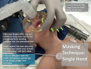
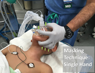
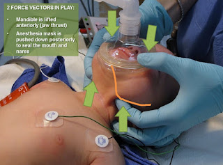
Masking Techniques in Pediatrics

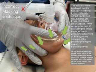
Masking Techniques in Pediatrics


Masking Techniques in Pediatrics

Masking Techniques in Pediatrics

Masking Techniques in Pediatrics

Masking Techniques in Pediatrics

Masking Techniques in Pediatrics

Masking Techniques in Pediatrics

Masking Techniques in Pediatrics

Masking Techniques in Pediatrics

Masking Techniques in Pediatrics

Masking Techniques in Pediatrics