Educational Blog about Anesthesia, Intensive care and Pain management

Goodpasture’s Syndrome

 Goodpasture’s Syndrome

Goodpasture’s Syndrome


-A rapidly progressive syndrome of glomerulonephritis and pulmonary hemorrhage, in association with antibodies to glomerular basement membrane (anti-GBM) type IV collagen, detectable in plasma by radioimmunoassay and by immunofluorescence techniques on muscle biopsy.

-These antibodies cross-react with the alveolar basement membrane, although those individuals with lung hemorrhage are usually smokers, and those with isolated anti-GBM nephritis are non-smokers.

-The term is often applied more loosely, to any disease with pulmonary hemorrhage and glomerulonephritis, whether or not anti-GBM antibodies are present.

-Systemic vasculitides such as PAN and Wegener’s granulomatosis cause lung hemorrhage and renal failure, but without the antibodies.

Preoperative Findings:

1. Patient presents with cough, dyspnea, hemoptysis (that can be massive), and anemia.

2. The pulmonary lesions proceed to interstitial fibrosis, with hemosiderin deposits. Lung function tests show a restrictive type of abnormality.

3. Glomerulonephritis, which usually follows or coincides with pulmonary lesions, may produce proteinuria, hematuria, and casts. The end result is renal failure.

4. Treatment may be with corticosteroids (prednisone) for pulmonary hemorrhage, cytotoxic drugs (cyclophosphamide) to stop renal damage, and plasmapheresis to remove antibodies.

Anesthetic Problems:

1. Poor respiratory function with hypoxemia and respiratory alkalosis.

2. Pulmonary hemorrhage that may, on occasions, be life-threatening.

3. Impaired renal function and sometimes renal failure.

4. Hypochromic anemia and a high ESR.

5. Patients may be receiving immunosuppressives or steroids, or undergoing plasma exchange, to reduce the antibody titer. Plasma exchange may reduce levels of plasma cholinesterase.

Anesthetic Management:

1. Preoperative assessment of lung function and, in particular, blood gases. Elective pulmonary surgery should not be undertaken during periods of active hemorrhage.

2. Assessment of renal function and appropriate management.

3. The management of a successful pregnancy has been described. It requires regular assessment of pulmonary and renal function. The use of steroids was associated with hyperglycemia, which needed treatment with insulin. Plasmapheresis was required.

Eisenmenger’s Syndrome

 Eisenmenger’s Syndrome

Eisenmenger’s Syndrome


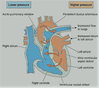
-A rare syndrome of pulmonary hypertension associated with a reversed or bidirectional cardiac shunt, occurring through a large communication between the left and right sides of the heart. The defect may be interventricular, interatrial, or aortopulmonary.

-The development of Eisenmenger’s syndrome, from the initial left to right shunt, is usually a gradual process. Contributory factors to pulmonary hypertension are hypoxia, high pulmonary blood flow, and high left atrial pressure.

-Irreversible structural changes take place in the small vessels, causing pulmonary vascular obstruction and a reduction in the size of the capillary bed. The pulmonary artery pressure is the same as or sometimes exceeds, the systemic arterial pressure.

-The incidence of this syndrome is decreasing because of the more vigorous approach to diagnosis and treatment of congenital heart disease in childhood.

Preoperative Findings:

1. Presenting symptoms include dyspnea, tiredness, episodes of cyanosis, syncope, or chest pain. Hemoptysis may occur.

2. The direction of the shunt, and hence the presence or absence of cyanosis, depends on several factors. These include hypoxemia, pulmonary and systemic pressure differences, and intravascular volume. It can also be affected by certain drugs.

3. Sleep studies have shown that there is a nocturnal deterioration in arterial oxygen saturation, which seems to be related to ventilation/perfusion distribution abnormalities occurring in the supine position.

4. Chest X-ray shows right ventricular hypertrophy, and ECG indicates varying degrees of right ventricular hypertrophy and strain.

5. Complications include thrombosis secondary to polycythemia, air embolus, bacterial endocarditis, gout, cholelithiasis, and hypertrophic osteoarthropathy. A cerebral abscess may occur secondary to clot embolism.

Anesthetic Problems:

1. Reductions in systemic arterial pressure by myocardial depression or loss of sympathetic tone are potentially dangerous. Reversal of the shunt may occur, and sudden death has been reported.

Hypovolemia and dehydration are poorly tolerated. Syntocinon may cause a dramatic reduction in SpO2 secondary to vasodilatation.

2. Sinus tachycardia results from exercise or emotion, and episodes of SVT are common after the age of 30. The onset of atrial fibrillation is associated with a sudden deterioration in the condition of the patient.

3. General anesthesia tends to be favored since the reduction in systemic vascular resistance associated with regional blockade increases the shunt. However, successful use of epidural anesthesia for bilateral inguinal herniorrhaphy, and Cesarean section, have been reported.

4. Pregnancy is contraindicated because it carries considerable risks. Recent maternal mortality rates of 40% have been reported. A cesarean section may increase it to over 60%. Termination of pregnancy is usually recommended in the first trimester but is still associated with a mortality of 7%.

5. Patients are at risk from paradoxical air or clot embolism, and infective endocarditis.

Anesthetic Management:

1. Understanding the pathophysiology of the complex is essential, and both pregnancy and non-cardiac surgery require a multidisciplinary approach.

2. Maintenance of an adequate circulating blood volume is important. Myocardial depressants and peripheral vasodilators should be used with caution. Bradycardia must be prevented. If regional anesthesia is used, the block should be instituted with caution, and hypovolemia avoided.

3. It is unclear whether oxygen can cause pulmonary vasodilatation. Although the pulmonary vascular resistance was believed to be fixed in pulmonary hypertension, a high oxygen concentration has been shown to reduce it during cesarean section.

4. Maintenance of systemic vascular resistance is critical. The use of a norepinephrine infusion before induction has been described. Alpha-adrenergic vasopressors, such as methoxamine or phenylephrine, have also been used for the treatment of hypotension on induction of anesthesia.

5. Pulmonary ventilation should be performed with low inflation pressures and early tracheal extubation is advised, because of the deleterious effects of IPPV.

6. Air must be completely eliminated from all intravenous lines and the epidural space should be located with loss of resistance to saline, not to air.

7. Antibiotic prophylaxis against bacterial endocarditis.

8. Low-dose heparin may reduce the risk of emboli.

9. Patients are usually advised against pregnancy. If anesthesia is required either for termination of pregnancy or operative delivery, intensive cardiac care is indicated. It has been suggested that those reaching the end of the second trimester should be admitted to the hospital until delivery and given heparin 20000-40000 units daily and oxygen therapy. Successful epidural anesthesia has been reported for cesarean section.

Tetralogy of Fallot

 Tetralogy of Fallot

Tetralogy of Fallot


-Tetralogy of Fallot (TOF) is a congenital cardiac abnormality. The primary defects are pulmonary infundibular stenosis and ventricular septal defect (VSD). The VSD is sufficiently large for the pressure in both ventricles to be equal to that of the aorta. The tetralogy is completed by two secondary features, a variable degree of overriding of the aorta, and right ventricular hypertrophy.

-Dynamic right ventricular outflow obstruction may occur (infundibular spasm), which is accentuated by sympathetic stimulation. The fraction of the right to left shunt depends primarily upon the relative resistances between the pulmonary (or right ventricular) and systemic outflows.

-If TOF is associated with patent foramen ovale (PFO) or atrial septal defect (ASD), it is a Pentalogy of Fallot.

-The aim of the surgery is to relieve the right ventricular outflow obstruction and to close the VSD. The traditional management by a two-stage repair has been replaced by definitive correction. Recent surgical advances include conduits to connect the RV to the PA and transatrial repair of the VSD.

The problems encountered during anesthesia will depend upon whether or not corrective surgery has been undertaken, and the functional result.

Preoperative Findings:

1. Dyspnea may occur on exertion and is hypoxia-related. Cyanosis and finger clubbing are variable, depending on the degree of pulmonary stenosis and the size of the shunt. Polycythemia (erythrocytosis) is common. There is a pulmonary stenotic murmur, but no murmur from the VSD because of the size of the defect. Squatting is thought to reduce the fraction of the shunt since kinking the large arteries increases systemic vascular resistance. Squatting is commonly seen in children with uncorrected lesions.

2. ECG shows right atrial and right ventricular hypertrophy, right axis deviation, and right bundle branch block.

3. Echocardiography shows right atrial and right ventricular hypertrophy, VSD, PFO or ASD, pulmonary hypertension, and overriding of the aorta.

4. Chest X-ray shows right ventricular hypertrophy and oligemic lungs. In the 2.6–6% of individuals who also have an absent pulmonary valve, aneurysmal dilatation of the pulmonary arteries may cause bronchial compression.

5. Initial surgery may have been undertaken to anastomose a systemic to a pulmonary artery, to improve the pulmonary blood flow, and reduce cyanosis. A definitive procedure is now more commonly undertaken in infancy.

6. In patients who have undergone shunt surgery without a definitive repair, there is chronic hypoxia and polycythemia. These patients have a high mortality, and an increased risk of bacterial endocarditis, thrombotic stroke, emboli, and intracerebral abscess.

7. In adults who have undergone repair there is an increased risk of arrhythmias, conduction defects, and sudden deaths, possibly related to mechanical events, such as ventricular dilatation and stretch, in the proximity of the conduction system. However, new approaches to surgery may result in a lower incidence of such problems in the future.

Anesthetic Problems:

1. In individuals with uncorrected lesions, the right to left shunt, and hence the cyanosis, is increased by a reduction in systemic vascular resistance produced by systemic vasodilatation. This may result from factors such as hypovolemia, drugs effect, or pyrexia.

2. Cyanosis is also worsened by an increase in pulmonary vascular resistance or spasm of the right ventricular infundibulum. Right ventricular outflow obstruction is due to increased contractility which is produced by increases in catecholamine output or the administration of drugs with positive inotropic effects. Anxiety, pain, hypercarbia, hypoxia, and acidosis are all precipitating factors. These cyanotic attacks or ‘tet’ spells, which can occur when awake or under anesthesia, may initiate a cycle of increasing hypoxia that can result in cerebral damage or death. Direct intraoperative observations of shunt direction and flow have been made with Doppler color flow imaging using epicardial leads. Patients with severe life-threatening hypoxemic spells, refractory to other treatment, responded to phenylephrine (5 mcg/kg plus an infusion of 0.4–2 mcg/kg/min).

3. Dehydration in the presence of polycythemia and high plasma viscosity may combine to increase the incidence of cerebral thrombosis. Polycythemia may also be associated with coagulation defects.

4. In patients with an absent pulmonary valve, positional airway compromise occurred secondary to bronchial compression of dilated pulmonary arteries.

5. A significant incidence of tracheal anomalies has been found.

6. In adults who have undergone repair, ventricular and atrial arrhythmias are common, particularly during exercise. It has been suggested that patients scheduled for elective surgery should have Holter monitors, or undergo exercise testing, in case antiarrhythmic treatment is needed first.

7. Cyanosed patients with Fallot’s rarely become pregnant. However, adults who have undergone corrective surgery are increasingly present during pregnancy with a favorable outcome.

Anesthetic Management:

1. Antibiotic prophylaxis against bacterial endocarditis.

2. A good premedication (morphine sulphate, or midazolam) to prevent excitement and anxiety.

3. In patients with cyanosis, measures are aimed at reducing the right to left shunt. Specific treatments of cyanotic attacks include:

a) Oxygen 100% to decrease PVR.

b) Pressor agents, such as phenylephrine (5-10 mcg/kg), to increase systemic vascular resistance.

c) Fluids to correct hypovolemia.

d) Propranolol (0.1 mg/kg) or Esmolol (0.5 mg/kg) to decrease outflow tract obstruction (decrease contractility and infundibular spasm).

e) Deepening of light anesthesia to reduce tachycardia associated with catecholamine output.

f) Compression of the femoral artery or the abdominal aorta against the vertebrae, sufficiently firmly to stop the femoral artery pulsations.

4. Techniques to avoid hypoxia and hypercarbia, and minimize vasodilatation and sudden increases in cardiac output. Ketamine, and Morphine sulphate (0.05-0.1 mg/kg), have been used.

5. Hydration is maintained in the perioperative period and, if there is severe polycythemia, venesection may be necessary.

6. Metabolic acidosis should be prevented or treated with sodium bicarbonate (1-2 mmol/kg).

7. In pregnancy, the outcome is satisfactory in patients whose ventricular function is good, and in whom no residual shunt occurs. However, close observation should be undertaken by an experienced team.

8. Venesection for erythrocytosis that is associated with cyanotic congenital heart disease should only take place if there are symptoms of hyperviscosity with a hematocrit >65%, and only provided volume replacement takes place at the same time.

9. Avoid muscle relaxants with histamine release e.g. atracurium, and use cisatracurium or rocuronium.

9. Mechanical ventilation:

-Increase FiO2

-Avoid increased airway pressure (< 15 cmH2O)

-Increase respiratory rate

-Avoid PEEP

Coarctation of Aorta

 Coarctation of Aorta

Coarctation of Aorta


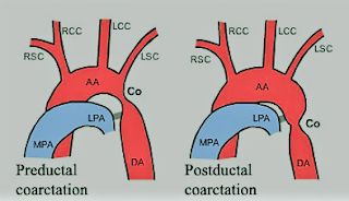
-A congenital narrowing of the aorta that may be pre-or post-ductal.

-The preductal form is usually a long, narrow segment, and is associated with other cardiac defects. This type generally presents with heart failure before the age of 1 year and requires treatment in a pediatric cardiac surgical unit.

-The postductal form, however, is often asymptomatic, and the patient may present in later life for surgery of some other condition, or for correction of the coarctation itself. Even after correction of coarctation, abnormalities can continue.

-Those who have undergone repair show persistent alterations in left ventricular function and left ventricular mass, together with resting gradients between the arm and leg. There is a higher incidence than normal of ischemic heart disease and sudden death.

-The optimal time for repair is disputed. Hypertension is more common in late correction, but repair at a young age is associated with a higher risk of recoarctation. In addition, persistent abnormalities in aortic stiffness have been found.

-Adult repairs are associated with a higher incidence of significant aortic valve disease (58%), compared with 37% of those repaired in childhood.

Preoperative Findings:

1. There may be moderate hypertension, the arm blood pressure being higher than that in the leg. If the left subclavian arises at or below the constriction there may be an absent or reduced left radial pulse. If both radial and femoral pulses are felt together, the small volume and delay of the femoral pulse will be obvious.

2. Collateral circulation develops in the internal mammary, intercostal, and subscapular arteries. The latter may be seen if the scapula is illuminated from the side.

3. A systolic murmur is usually heard along the left sternal edge radiating up into the neck.

4. Chest X-ray may show notching of the undersides of the ribs, secondary to intercostal arteries dilatation. There may be pre-and post-stenotic dilatation of the aorta.

5. Occasionally, cerebral berry aneurysms coexist with coarctation. In such cases, the high arterial pressure increases the risk of subarachnoid hemorrhage.

6. There is a 25–50% incidence of the bicuspid aortic valve and some aortic regurgitation.

7. Unusually, angina or left ventricular failure may present late in untreated adult coarctation.

8. Patients may require balloon dilatation or surgical resection.

Anesthetic Problems:

1. Before elective surgery, if undiagnosed coarctation is found, treatment of the coarctation may be considered to be the priority. Even if coarctation has been treated, the possibility of residual cardiovascular abnormalities should be considered, since there is an increased risk of premature death compared with the normal population. Causes include aneurysms (cerebral, at the operative site, other parts of the aorta and intercostal arteries), hypertension, myocardial infarction, and cardiac failure. Balloon angioplasty may carry less risk.

2. Patients may present with hypertension-related complications.

3. Any operation in the area of the dilated collateral vessels may result in heavy bleeding, especially when the chest is opened.

4. Hypoperfusion of the spinal cord. This may cause paraplegia, and is more likely in those patients with few collaterals. Induced hypotension for clipping of cerebral aneurysms may compromise spinal cord perfusion.

5. Susceptibility to bacterial endocarditis.

6. If there are left subclavian abnormalities, the left arm cannot be used for blood pressure monitoring.

7. Patients may present during pregnancy with hypertension, which may be confused with preeclampsia.

Anesthetic Management:

1. Antihypertensive therapy should be used until the day of operation. Beta-adrenoceptor blockers may reduce the hypertensive response to intubation.

2. Antibiotic prophylaxis against bacterial endocarditis.

3. During clipping of an intracranial aneurysm, monitoring of the femoral artery pressure should be undertaken to assess spinal cord perfusion during induced hypotension. A mean distal aortic pressure above 50 mmHg has been suggested as adequate for spinal cord perfusion.

Hydatid Disease

 Hydatid Disease

Hydatid Cyst


-Hydatid cysts are the larval stage of the tapeworm, Echinococcus granulosus.

-Dogs are the main hosts. Man and sheep are intermediate hosts.

-Hydatid disease is not uncommon amongst the mid-Wales farming communities, and up to 26% of farm dogs in this area have E. granulosus.

-The incidence is also high in some regions of the Mediterranean, North Africa, the Middle East, and Australia.

-If the ova are ingested by man, embryos are released when the chitinous coat is digested.

-These enter the liver by the portal vein. They may be destroyed, or they may develop into a cyst.

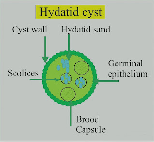
-In man, the cysts are found in the liver (65%), lung (25%), muscles (5%), bone (3%), and brain (1%). Each cyst is two-layered and contains straw-colored fluid in which there are free scolices, brood capsules containing scolices, and daughter cysts.

-Around the cyst is an area of compressed host tissue and fibrosis known as the pericyst. In 5–10% of cases, the cyst will die, and calcification may occur.

Preoperative Findings:

1. Hepatic cysts occur most frequently in the right lobe. A bacterial infection may result in a liver abscess. Rupture into a bile duct, or bile duct obstruction can occur and produce biliary colic. There may be jaundice. The number and location of the cysts are shown on a CT scan or ultrasound.

2. Pulmonary cysts can present with hemoptysis, dyspnea, cough, or chest pain. Chest X-ray may show a variety of appearances including an oval opacity, evidence of bronchial fistula formation, or rupture of the cyst with the development of a fluid level.

3. Eosinophilia occurs in about 30% of cases. The Casoni skin test is still used for screening. Immunoelectrophoresis is the most specific test. Complement fixation test is positive in up to 80% of cases. The hemagglutination test detects a specific antibody.

Anesthetic Problems:

1. Pulmonary hydatid cysts can cause bronchial obstruction, and occasionally they may rupture into the airway. If this happens, flooding of the lungs occurs, with widespread dissemination of the scolices.

2. Hydatid fluid is highly antigenic, and rupture of a cyst has occasionally produced sudden death from an anaphylactic reaction. Anaphylaxis to hydatid was confirmed by increased serum histamine and tryptase levels, with elevated levels of total and echinococcus-specific IgE antibodies.

3. Patients can develop anaphylaxis of unknown origin as interventricular hydatid cystic mass.

4. Cerebral cysts can cause increased intracranial pressure.

5. Scolicidal agents are potentially toxic and their use in combination with surgery may increase the complication rate.

6. Postoperative complications following surgery for hydatid cyst of the lung included prolonged air leak and aspiration pneumonia.

Anesthetic Management:

1. Surgical removal is indicated, except in older patients with small cysts. Meticulous care must be taken to avoid the rupture and spread of the fertile scolices.

2. Relatively new drugs, such as mebendazole and albendazole, are being tested as scolicidal agents. However, there is no evidence that they are effective in the treatment of pulmonary hydatid.

3. Pulmonary cysts. Protective formalin soaked packs are placed around the wound, an incision is made through the pericyst and the cyst is carefully extruded by the anesthetist, using gentle hand ventilation.

4. Because of the risk of an anaphylactic reaction, epinephrine (adrenaline), metaraminol, isoprenaline, and steroids must be immediately available.

Gilbert’s Disease

Gilbert’s Disease (Idiopathic Unconjugated Hyperbilirubinemia)

Gilbert’s Disease


-An autosomal dominant, benign condition, in which there is a mildly elevated unconjugated bilirubin, without either structural liver disease or hemolytic anemia.

-It possibly results from an impaired uptake and conjugation of bilirubin, secondary to a deficiency of UDP glucuronyl transferase.

-The diagnosis is made on a level of unconjugated bilirubin of 17–102 mmol/l, alternatively, a serum total bilirubin of 25–50 mmol/l after a 24-h restricted diet.

Preoperative abnormalities:

1. Serum unconjugated bilirubin is increased, but usually to a level <50 mmol/l and clinical jaundice is barely detectable. However, fluctuating mild jaundice may occur, particularly in the presence of stress, infection, starvation, or surgery.

2. Other liver function tests are normal, and there is no hemolytic anemia.

Anesthetic Problems:

1. The condition itself is of no significance. However, the appearance of jaundice postoperatively may suggest more serious problems, therefore the confirmation of Gilbert’s syndrome as the cause is useful.

2. Starvation may elevate the bilirubin level and produce visible jaundice.

3. The metabolism of morphine, and papaveretum, may be delayed.

4. There are potential toxicological implications for any drug or chemical that is eliminated primarily via glucuronidation mechanisms such as paracetamol.

Anesthetic Management:

1. If Gilbert’s disease is suspected, the administration of nicotinic acid 50 mg IV will double or triple the plasma unconjugated bilirubin within 3 h. In normal patients, or in those with other liver diseases, the increase will be less.

2. Morphine, papaveretum, and paracetamol should be used with caution.

3. Early morning surgery and a dextrose infusion will reduce the increase in bilirubin provoked by starvation.

Budd–Chiari Syndrome

 Budd–Chiari Syndrome

Budd–Chiari Syndrome


-A syndrome caused by obstruction to the hepatic venous outflow and resulting in a clinical picture of hepatomegaly and portal hypertension. It may be secondary to hematological disorders, malignancy, oral contraceptives, heart failure, or constrictive pericarditis.

-The three main sites of obstruction are:

1-Inferior vena cava

2-Large hepatic veins

3-Small intrahepatic venules.

-The condition may be acute or chronic. Acute Budd–Chiari syndrome with hepatocyte necrosis may require urgent portosystemic, decompressive, surgical, or interventional radiological procedures.

-Untreated hepatic venous thrombosis usually results in progressive liver failure and death. Medical treatment is of little help, therefore shunt procedures or liver transplantation will be needed.

Preoperative Findings:

1. Abdominal pain and swelling are the commonest symptoms. Ascites and hepatomegaly will be present in the majority. Vomiting, splenomegaly, jaundice, or bleeding from esophageal varices may also occur. If the vena cava is involved there will be dependent edema.

2. Liver function abnormalities depend on the site and severity of the obstruction. Liver biopsy may show outflow obstruction, hepatic necrosis, and fibrosis.

3. Etiological factors include polycythemia, paroxysmal nocturnal hemoglobinuria, antiphospholipid syndrome, protein C deficiency, factor V Leiden, myeloproliferative disorders, and mechanical factors, such as webs and tumors. Hepatic venous thrombosis has been reported in association with ulcerative colitis, but in the patient described, iron deficiency had concealed an underlying polycythemia vera.

4. Treatment is aimed at the preservation of liver function and includes thrombolysis, angioplasty, stent placement, portacaval shunt, mesoatrial shunt, and liver transplantation.

Anesthetic Problems:

1. Hepatic function may be compromised.

2. Surgery may be required for portacaval or mesoatrial shunt or liver transplantation. This has been described in a patient with paroxysmal nocturnal hemoglobinuria.

3. Patients may present during pregnancy when the condition must be distinguished from HELLP syndrome and acute fatty necrosis of the liver. Antiphospholipid syndrome and preeclampsia in a primipara presented with Budd–Chiari syndrome from thrombosis of the right hepatic veins.

Thrombotic thrombocytopenic purpura resulted in postpartum hepatic venous thrombosis. Paroxysmal nocturnal hemoglobinuria has been associated with hemolytic crises and Budd–Chiari syndrome during pregnancy. Maternal deaths have been reported.

Anesthetic Management:

1. Hematological examination is important because, in the absence of mechanical causes for hepatic vein thrombosis, there is a high incidence of underlying hematological abnormalities; these include myeloproliferative disorders, and paroxysmal nocturnal hemoglobinuria, systemic lupus erythematosus, and antithrombin III deficiency.

2. Assessment of liver function, including coagulation.

3. Low-dose heparin infusion from the first postoperative day has been recommended, with subsequent low-dose aspirin and long-term anticoagulation to reduce the risks of graft thrombosis.

4. Liver transplantation will cure the inherited thrombophilias, for example, factor V Leiden, protein C and S, etc.

5. In a patient with paroxysmal nocturnal hemoglobinuria who developed Budd–Chiari syndrome, the resolution was achieved following bone marrow transplantation.

6. Pregnant patients with paroxysmal nocturnal hemoglobinuria should be carefully monitored for the onset of Budd–Chiari syndrome.

Chiari Malformations

 Chiari Malformations

Chiari Malformations


-The Chiari malformation is a term encompassing a range of hindbrain maldevelopments.

-Problems include underdevelopment of the cranial fossa and overcrowding of the normally developed hindbrain, the risks of descent of hindbrain structures below the foramen magnum, and intermittent obstruction to CSF outflow from the fourth ventricle.

-There may be a relatively high CSF pressure in the head and a relatively lower one in the spine.

Types of Chiari Malformations:

There are four types.

Type I:

-Downward herniation of cerebellar tonsils of at least 3–5 mm below the foramen magnum and hindbrain overcrowding results in direct compression of the tissue.

-Clinical symptoms thus relate to CSF disturbances and direct compression of nervous tissue; they include headaches, pseudotumor-like episodes, a syndrome resembling Meniere’s disease, lower cranial nerve signs, and spinal cord disturbances without syringomyelia.

-It is mostly seen in young adults.

Type II (Arnold–Chiari):

-It is the most important and includes; thoracolumbar myelomeningocele, hypoplasia of the posterior fossa, and displacement of the cerebellar vermis, brainstem, and fourth ventricle.

-It presents in infancy or early childhood.

Type III: rare

-Dilated the fourth ventricle with cervical meningomyelocele.

Type IV: rare

-Cerebellar hypoplasia.

Other abnormalities:

1. May be associated with syringomyelia or syringobulbia, other skeletal abnormalities of the skull base and cervical vertebrae, or myelomeningocele. Syringomyelia is the term for an expanding, longitudinal cystic cavity within the spinal cord. In the communicating variety, there is continuity between the syrinx and the CSF in the central canal.

2. May need decompression of foramen magnum or cervical spine.

3. Progressive hydrocephalus may occur which needs shunt surgery.

4. Obstructive sleep apnea.

5. Recurrent pulmonary aspiration.

6. May exhibit abnormal autonomic control.

7. Patients may present with respiratory arrest.

8. Sudden unexpected death may occur.

Anesthetic Problems:

1. Increased ICP may lead to coning.

2. Symptoms may start after a dural puncture.

3. Bolus injections into the epidural space may lead to ICP increases in susceptible individuals.

4. Sleep apnea may occur in Chiari type I.

5. Acute cardiovascular collapse secondary to inadvertent brainstem compression in an infant.

Anesthetic Management:

1. A careful history should be taken in a patient with a ventriculoperitoneal shunt, to check for any signs that it is nonfunctioning. If there is doubt, referral to a neurosurgeon is indicated. This is particularly important if the notes are absent, there is poor history or lack of follow-up.

2. Try to prevent anything likely to increase ICP, such as coughing, or hypercarbia from heavy sedation or analgesia.

3. Avoid straining in labor. Epidural anesthesia for Cesarean section was performed in a patient with type I Chiari who had a previous difficult tracheal intubation. Small doses of local anesthetic should be titrated gradually, to avoid sudden distension of the epidural space or decreases in arterial pressure.

4. Spinal anesthesia should be avoided.

Ludwig’s Angina

Ludwig’s Angina

Ludwig’s Angina

Definition and Causes:

A rapidly spreading cellulitis of the floor of the mouth that can be produced by any infection. It involves the submandibular, sublingual, and submental spaces.

-Gram-positive cocci (usually streptococci), Staphylococcus aureus, and Staphylococcus epidermidis are now the most common organisms, but sometimes gram-negative rods or anaerobes are responsible. In 50% of cases, more than one organism is isolated.

-It is frequently precipitated by a dental infection involving the second and third lower molars, but trauma may be contributory.

-The condition usually arises from the teeth, but tongue piercing with metal barbells has provided a novel source of infection.

-The frequency is generally decreasing, but there is now a higher incidence in patients with associated systemic diseases. Antibiotics and aggressive surgical treatment have dramatically improved the mortality rate.

-Airway management is controversial, airway compromise develops insidiously, but the actual obstruction is abrupt.

Preoperative Findings:

1. Bilateral submandibular swelling proceeds to brawny induration of the neck. Although the submandibular space is primarily involved in Ludwig’s angina, spread into adjacent fascial spaces may occur.

2. Elevation of the tongue caused by cellulitis of the floor of the mouth.

3. Dysphagia secondary to swelling, and trismus.

4. Gradual onset, or sudden, upper airway obstruction.

5. There is frequently an underlying systemic condition, such as DM, AIDS, or substance abuse.

6. Other complications include; bacteremia, aspiration, retropharyngeal abscess, empyema, mediastinitis, internal jugular vein thrombosis, and pericarditis.

7. Fever, leukocytosis, and increased ESR.

Anesthetic Problems:

1. Trismus, not necessarily relieved by muscle relaxants, may make oral intubation difficult or impossible.

2. Sudden total upper airway obstruction producing hypoxia.

3. Intravenous induction of anesthesia may be hazardous because it can result in apnea and an inability to maintain ventilation on a mask.

4. On a prolonged history of dental sepsis, sepsis may track down through the retropharyngeal space into the posterior mediastinum, requiring awake oral intubation, ventilatory support, and an eventual thoracotomy for a thoracic empyema.

Anesthetic Management:

1. Aggressive early treatment with antibiotics reduces airway problems and the need for surgical intervention.

2. Less severely affected patients may be managed by close observation, but only provided that staff is available who can manage acute obstruction. Some argue the case for early tracheostomy.

3. Surgical drainage with or without tooth extraction.

4. Anesthesia for surgery in which there is trismus, but without compromise of the upper airway:

a) Awake fibreoptic nasal intubation but with facilities for emergency tracheostomy available.

b) Inhalation induction and laryngoscopy. If the trismus relaxes and the vocal cords can be seen, a neuromuscular blocker can be given.

c) Tracheostomy may be required in the presence of spreading edema.

5. Airway maintenance in the compromised airway:

-Signs of airway obstruction including; stridor, dyspnea, dysphagia, secretions, and deteriorating oxygen saturations, may indicate the need for rapid, active intervention. In these patients, sedative premedication should be avoided and a drying agent is given.

-If there is significant stridor, a tracheostomy under local anesthesia may be considered.

-Emergency cricothyroidotomy under local anesthesia has also been reported.