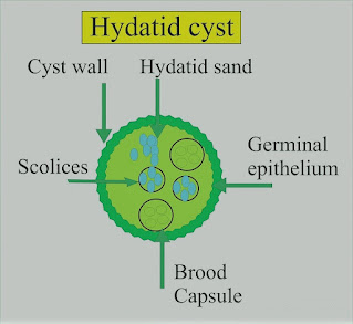
Hydatid Disease
-Hydatid cysts are the larval stage
of the tapeworm, Echinococcus granulosus.
-Dogs are the main hosts. Man and
sheep are intermediate hosts.
-Hydatid disease is not uncommon
amongst the mid-Wales farming communities, and up to 26% of farm dogs in this
area have E. granulosus.
-The incidence is also high in some
regions of the Mediterranean, North Africa, the Middle East, and Australia.
-If the ova are ingested by man,
embryos are released when the chitinous coat is digested.
-These enter the liver by the portal
vein. They may be destroyed, or they may develop into a cyst.
-In man, the cysts are found in the
liver (65%), lung (25%), muscles (5%), bone (3%), and brain (1%). Each cyst is
two-layered and contains straw-colored fluid in which there are free scolices,
brood capsules containing scolices, and daughter cysts.
-Around the cyst is an area of
compressed host tissue and fibrosis known as the pericyst. In 5–10% of cases, the cyst will die, and calcification may occur.
Preoperative Findings:
1. Hepatic cysts occur most
frequently in the right lobe. A bacterial infection may result in a liver
abscess. Rupture into a bile duct, or bile duct obstruction can occur and
produce biliary colic. There may be jaundice. The number and location of the
cysts are shown on a CT scan or ultrasound.
2. Pulmonary cysts can present with
hemoptysis, dyspnea, cough, or chest pain. Chest X-ray may show a variety of
appearances including an oval opacity, evidence of bronchial fistula formation,
or rupture of the cyst with the development of a fluid level.
3. Eosinophilia occurs in about 30%
of cases. The Casoni skin test is still used for screening.
Immunoelectrophoresis is the most specific test. The complement fixation test is positive
in up to 80% of cases. The hemagglutination test detects a specific antibody.
Anesthetic Problems:
1. Pulmonary hydatid cysts can cause
bronchial obstruction, and occasionally they may rupture into the airway. If
this happens, flooding of the lungs occurs, with widespread dissemination of
the scolices.
2. Hydatid fluid is highly
antigenic, and the rupture of a cyst has occasionally produced sudden death from an
anaphylactic reaction. Anaphylaxis to hydatid was confirmed by increased serum
histamine and tryptase levels, with elevated levels of total and echinococcus-specific
IgE antibodies.
3. Patients can develop anaphylaxis
of unknown origin as interventricular hydatid cystic mass.
4. Cerebral cysts can cause
increased intracranial pressure.
5. Scolicidal agents are potentially
toxic and their use in combination with surgery may increase the complication
rate.
6. Postoperative complications
following surgery for hydatid cyst of the lung included prolonged air leak and
aspiration pneumonia.
Anesthetic Management:
1. Surgical removal is indicated,
except in older patients with small cysts. Meticulous care must be taken to
avoid the rupture and spread of the fertile scolices.
2. Relatively new drugs, such as
mebendazole and albendazole, are being tested as scolicidal agents. However,
there is no evidence that they are effective in the treatment of pulmonary
hydatid.
3. Pulmonary cysts. Protective
formalin-soaked packs are placed around the wound, an incision is made through
the pericyst, and the cyst is carefully extruded by the anesthetist, using
gentle hand ventilation.
4. Because of the risk of an anaphylactic reaction, epinephrine (adrenaline), metaraminol, isoprenaline, and steroids must be immediately available.


