Educational Blog about Anesthesia, Intensive care and Pain management

Gilbert’s Disease

Gilbert’s Disease (Idiopathic Unconjugated Hyperbilirubinemia)

Gilbert’s Disease


-An autosomal dominant, benign condition, in which there is a mildly elevated unconjugated bilirubin, without either structural liver disease or hemolytic anemia.

-It possibly results from an impaired uptake and conjugation of bilirubin, secondary to a deficiency of UDP glucuronyl transferase.

-The diagnosis is made on a level of unconjugated bilirubin of 17–102 mmol/l, alternatively, a serum total bilirubin of 25–50 mmol/l after a 24-h restricted diet.

Preoperative abnormalities:

1. Serum unconjugated bilirubin is increased, but usually to a level <50 mmol/l, and clinical jaundice is barely detectable. However, fluctuating mild jaundice may occur, particularly in the presence of stress, infection, starvation, or surgery.

2. Other liver function tests are normal, and there is no hemolytic anemia.

Anesthetic Problems:

1. The condition itself is of no significance. However, the appearance of jaundice postoperatively may suggest more serious problems, therefore the confirmation of Gilbert’s syndrome as the cause is useful.

2. Starvation may elevate the bilirubin level and produce visible jaundice.

3. The metabolism of morphine, and papaveretum, may be delayed.

4. There are potential toxicological implications for any drug or chemical that is eliminated primarily via glucuronidation mechanisms such as paracetamol.

Anesthetic Management:

1. If Gilbert’s disease is suspected, the administration of nicotinic acid 50 mg IV will double or triple the plasma unconjugated bilirubin within 3 h. In normal patients, or in those with other liver diseases, the increase will be less.

2. Morphine, papaveretum, and paracetamol should be used with caution.

3. Early morning surgery and a dextrose infusion will reduce the increase in bilirubin provoked by starvation.

Hydatid Disease

 Hydatid Disease

Hydatid Cyst


-Hydatid cysts are the larval stage of the tapeworm, Echinococcus granulosus.

-Dogs are the main hosts. Man and sheep are intermediate hosts.

-Hydatid disease is not uncommon amongst the mid-Wales farming communities, and up to 26% of farm dogs in this area have E. granulosus.

-The incidence is also high in some regions of the Mediterranean, North Africa, the Middle East, and Australia.

-If the ova are ingested by man, embryos are released when the chitinous coat is digested.

-These enter the liver by the portal vein. They may be destroyed, or they may develop into a cyst.

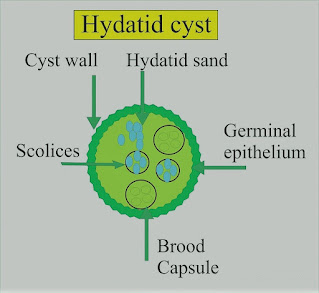
-In man, the cysts are found in the liver (65%), lung (25%), muscles (5%), bone (3%), and brain (1%). Each cyst is two-layered and contains straw-colored fluid in which there are free scolices, brood capsules containing scolices, and daughter cysts.

-Around the cyst is an area of compressed host tissue and fibrosis known as the pericyst. In 5–10% of cases, the cyst will die, and calcification may occur.

Preoperative Findings:

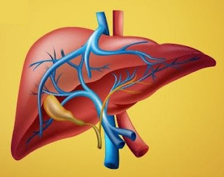
1. Hepatic cysts occur most frequently in the right lobe. A bacterial infection may result in a liver abscess. Rupture into a bile duct, or bile duct obstruction can occur and produce biliary colic. There may be jaundice. The number and location of the cysts are shown on a CT scan or ultrasound.

2. Pulmonary cysts can present with hemoptysis, dyspnea, cough, or chest pain. Chest X-ray may show a variety of appearances including an oval opacity, evidence of bronchial fistula formation, or rupture of the cyst with the development of a fluid level.

3. Eosinophilia occurs in about 30% of cases. The Casoni skin test is still used for screening. Immunoelectrophoresis is the most specific test. The complement fixation test is positive in up to 80% of cases. The hemagglutination test detects a specific antibody.

Anesthetic Problems:

1. Pulmonary hydatid cysts can cause bronchial obstruction, and occasionally they may rupture into the airway. If this happens, flooding of the lungs occurs, with widespread dissemination of the scolices.

2. Hydatid fluid is highly antigenic, and the rupture of a cyst has occasionally produced sudden death from an anaphylactic reaction. Anaphylaxis to hydatid was confirmed by increased serum histamine and tryptase levels, with elevated levels of total and echinococcus-specific IgE antibodies.

3. Patients can develop anaphylaxis of unknown origin as interventricular hydatid cystic mass.

4. Cerebral cysts can cause increased intracranial pressure.

5. Scolicidal agents are potentially toxic and their use in combination with surgery may increase the complication rate.

6. Postoperative complications following surgery for hydatid cyst of the lung included prolonged air leak and aspiration pneumonia.

Anesthetic Management:

1. Surgical removal is indicated, except in older patients with small cysts. Meticulous care must be taken to avoid the rupture and spread of the fertile scolices.

2. Relatively new drugs, such as mebendazole and albendazole, are being tested as scolicidal agents. However, there is no evidence that they are effective in the treatment of pulmonary hydatid.

3. Pulmonary cysts. Protective formalin-soaked packs are placed around the wound, an incision is made through the pericyst, and the cyst is carefully extruded by the anesthetist, using gentle hand ventilation.

4. Because of the risk of an anaphylactic reaction, epinephrine (adrenaline), metaraminol, isoprenaline, and steroids must be immediately available.

Coarctation of Aorta

 Coarctation of Aorta

Coarctation of Aorta


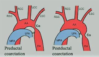
-A congenital narrowing of the aorta that may be pre- or post-ductal.

-The preductal form is usually a long, narrow segment, and is associated with other cardiac defects. This type generally presents with heart failure before the age of 1 year and requires treatment in a pediatric cardiac surgical unit.

-The postductal form, however, is often asymptomatic, and the patient may present in later life for surgery of some other condition, or for correction of the coarctation itself. Even after correction of coarctation, abnormalities can continue.

-Those who have undergone repair show persistent alterations in left ventricular function and left ventricular mass, together with resting gradients between the arm and leg. There is a higher incidence than normal of ischemic heart disease and sudden death.

-The optimal time for repair is disputed. Hypertension is more common in late correction, but repair at a young age is associated with a higher risk of recoarctation. In addition, persistent abnormalities in aortic stiffness have been found.

-Adult repairs are associated with a higher incidence of significant aortic valve disease (58%), compared with 37% of those repaired in childhood.

Preoperative Findings:

1. There may be moderate hypertension, the arm blood pressure being higher than that in the leg. If the left subclavian arises at or below the constriction there may be an absent or reduced left radial pulse. If both radial and femoral pulses are felt together, the small volume and delay of the femoral pulse will be obvious.

2. Collateral circulation develops in the internal mammary, intercostal, and subscapular arteries. The latter may be seen if the scapula is illuminated from the side.

3. A systolic murmur is usually heard along the left sternal edge radiating up into the neck.

4. Chest X-ray may show notching of the undersides of the ribs, secondary to intercostal arteries dilatation. There may be pre-and post-stenotic dilatation of the aorta.

5. Occasionally, cerebral berry aneurysms coexist with coarctation. In such cases, the high arterial pressure increases the risk of subarachnoid hemorrhage.

6. There is a 25–50% incidence of the bicuspid aortic valve and some aortic regurgitation.

7. Unusually, angina or left ventricular failure may present late in untreated adult coarctation.

8. Patients may require balloon dilatation or surgical resection.

Anesthetic Problems:

1. Before elective surgery, if undiagnosed coarctation is found, treatment of the coarctation may be considered to be the priority. Even if coarctation has been treated, the possibility of residual cardiovascular abnormalities should be considered, since there is an increased risk of premature death compared with the normal population. Causes include aneurysms (cerebral, at the operative site, other parts of the aorta and intercostal arteries), hypertension, myocardial infarction, and cardiac failure. Balloon angioplasty may carry less risk.

2. Patients may present with hypertension-related complications.

3. Any operation in the area of the dilated collateral vessels may result in heavy bleeding, especially when the chest is opened.

4. Hypoperfusion of the spinal cord. This may cause paraplegia, and is more likely in those patients with few collaterals. Induced hypotension for clipping of cerebral aneurysms may compromise spinal cord perfusion.

5. Susceptibility to bacterial endocarditis.

6. If there are left subclavian abnormalities, the left arm cannot be used for blood pressure monitoring.

7. Patients may present during pregnancy with hypertension, which may be confused with preeclampsia.

Anesthetic Management:

1. Antihypertensive therapy should be used until the day of operation. Beta-adrenoceptor blockers may reduce the hypertensive response to intubation.

2. Antibiotic prophylaxis against bacterial endocarditis.

3. During clipping of an intracranial aneurysm, monitoring of the femoral artery pressure should be undertaken to assess spinal cord perfusion during induced hypotension. A mean distal aortic pressure above 50 mmHg has been suggested as adequate for spinal cord perfusion.