Educational Blog about Anesthesia, Intensive care and Pain management

Showing posts with label Regional anesthesia. Show all posts

Accidental Total Spinal Anesthesia

Accidental Total Spinal Anesthesia


Accidental Total Spinal Anesthesia

Definition:

➧ A syndrome of the central neurological blockade. 

➧ It occurs when a volume of local anesthetic (LA) solution, intended for epidural anesthesia, enters the subarachnoid space and ascends to the cervical region.

Causes: 

a) After a known dural tap: 

➧ Accidental total spinal analgesia may occur in association with the original epidural after a known dural tap. 

b) After a top-up dose: 

➧ Following a top-up dose, as a result of accidental puncture of the dura by the epidural catheter. 

➧ High spinal anesthesia has occurred after top-ups of epidural catheters. 

➧ This is unlikely to result from catheter migration but may happen as a result of accidental puncture of the dura by the epidural catheter. 

➧ If part of the catheter lies within the epidural space and part within the subarachnoid, with a slow injection of LA, the solution will emerge from the proximal holes, and with a rapid one from the more distal. 

Presentation: 

1-Unexpected rapid rise in the sensory level. 

2-Numbness or weakness in the upper extremities. 

3-There is severe hypotension and bradycardia secondary to blockade of the sympathetic outflow. Occasionally cardiac arrest occurs. 

4-Rapidly increasing paralysis involves the respiratory muscles, due to phrenic nerve paralysis, resulting in apnea and hypoxia. 

5-The pupils become dilated and consciousness is lost. 

6-Cardiovascular collapse usually takes place immediately after the injection, although delays of up to 45 min. have been reported. 

7-Deaths have occasionally been reported. 

Duration: 

➧ Apnea may vary from 20 min. to 6 h, unconsciousness from 25 min. to 4 h, while full recovery of sensation may take up to 9 h. 

➧ The lengths of time vary with the agent, the dose, and the volume of LA given. Bupivacaine lasts longer than lidocaine. 

Management: 

a) Precautions: 

1-A test dose of LA is recommended. 

2-The injection of 3 ml of LA containing epinephrine 1:200 000, followed by an adequate pause to assess the effects, has been suggested. 

3-It has been recommended that, if a dural puncture occurs during active labor when a cesarean section is required, then further attempts should not be made. Either a spinal or a general anesthetic should be employed as an alternative. 

b) If Total Spinal a. occurs: 

1-Call for help. 

2-A non-pregnant patient should be turned supine, and the legs elevated to encourage venous return. The pregnant patient should be tilted in the lateral position to prevent aortocaval compression

3-The lungs should be inflated with oxygen

4-A tracheal tube can then be inserted. IPPV may have to be continued for up to 2 h, depending upon the LA and the volume used

5-Intravenous fluids should be infused rapidly

6-A vasopressor agent such as Ephedrine IV in 5–10 mg increments up to 30 mg is recommended. Epinephrine (adrenaline) 0.1–0.5 mg may occasionally be required but should preferably be avoided in patients in labor

7-Atropine for bradycardia, use epinephrine if the patient is unstable

Failed Epidural Block

Failed Epidural Block


Introduction:

➧ Failure of epidural anesthesia and analgesia occurs in up to 30% of clinical practices. 

➧ Some technical factors can help to increase the primary and secondary success rates.

A) Technical factors influencing block success:

1-Patient Position:

➧ Patient positioning potentially affects needle placement by changing the relationship between osseous and soft tissues. 

a) Sitting Position 

➧ Results in shorter insertion times and higher accuracy at the first attempt than in the lateral position. 

➧ Causes more vagal reflexes than lateral position. 

➧ This leads to epidural venous plexus distension, which may theoretically increase the risk of vascular puncture, especially in parturients. 

b) Lateral Position 

➧ Increases the distance from the skin to the epidural space. 

➧ Results in more technical difficulties compared with the sitting position.

2-Puncture Site:

➧ Inaccurate dermatomal block-level or anatomical landmarks of neuraxial puncture are not suitable for the type of surgery.

3-Approach:

a) Midline Approach 

➧ Results in a higher incidence of paresthesia than the paramedian approach. 

➧ Results in a bloody puncture in non-pregnant adults than paramedian approach. 

➧ The ligamentum flavum is not continuous in all patients, and the presence of midline gaps may make the loss of resistance (LoR) to needle advancement and injection of air/saline less perceptible when the midline approach is used. 

b) Paramedian Approach 

➧ Results in faster catheter insertion times, and less dependent upon spine flexion. 

➧ Paramedian catheters cause less epidural tenting, and pass cephalad more reliably than midline catheters.

4-Identification of the Epidural Space:

➧ Correct placement obviously requires correct identification of the epidural space. The LoR using saline has become the most widely used method, while LoR to air and the hanging drop technique is less widely used with no difference in the success rate or adverse events, other than a 1.5% reduction in post-dural puncture headache [1] when using saline. 

➧ In obstetric epidurals, using saline for LoR results in fewer attempts than using air, but with comparable final success rates. 

➧ The use of the ‘preferred technique’ (i.e. the technique used by anesthetists 70% of the time) results in significantly fewer attempts, a lower incidence of paresthesia, and fewer dural punctures, irrespective of whether saline or air is used for LoR. 

➧ The hanging drop technique depends on negative pressure within the epidural space and is useful only in the sitting position. 

➧ Identification of the epidural space was reported at 2 mm deeper for the hanging drop when compared with LoR, possibly indicating an increased risk of dural puncture.

➧ Ultrasound pre-assessment of lumbar epidural space depth has been shown to correlate well with actual puncture depth in obese parturients. 

➧ The use of ultrasound led to less bony contact, a shorter time to block success, and decreased supplemental opioid requirements.

5-Epidural Catheter Location:

➧ Epidural catheters may primarily be placed incorrectly, or become dislodged during operation. 

➧ Primary misplacement of epidural catheters in the paravertebral space, in the pleural cavity, or intravascularly. 

➧ Transforaminal migration of the catheter tip and asymmetric spread during epidural analgesia. 

➧ During normal patient movement, epidural catheters may be displaced by centimeters. 

➧ Changes in epidural pressure and cerebrospinal fluid (CSF) oscillations can contribute to the displacement of epidural catheters. 

➧ Midline fat pedicles may form a barrier to the spread of local anesthetics (LA).

6-Catheter Insertion and Fixation:

➧ The catheter should be inserted at least 4 cm into the epidural space.

➧ Suturing of the epidural catheter is associated with less migration, but at the cost of increased inflammation at the puncture site. 

➧ Tunneling the epidural catheter for 5 cm is associated with less movement of the catheter and decreases catheter migration but it will not maintain the original position. 

➧ Tunneling caudal epidural catheter in children reduces the risk of bacterial colonization to levels comparable to untunneled lumbar catheters because tunneling places the catheter entry point above the diaper in babies and toddlers. 

➧ For lumbar and epidural catheters, the advantages of tunneling are less obvious and the need to prevent dislodgement must be weighed against the increased incidence of erythema at the puncture site, potentially linked to increased risk of bacterial colonization.

➧ Catheter fixation devices are available which may significantly reduce migration percentage and reduce rates of analgesic failure.

7-Test Dose:

➧ A test dose is given with two main objectives of detecting intrathecal or intravascular catheter placement. 

➧ A test dose of lidocaine (to detect intrathecal placement) and epinephrine (to detect intravascular placement) is recommended in patients without contraindications to epinephrine. 

➧ Specific regimens to detect intravascular catheter position: 

-Fixed epinephrine test dose for non-pregnant adult patients. 

-Fentanyl test dose for parturients. 

-Weight-adjusted epinephrine test dose for children. 

➧ Patients sensitive to intravascular epinephrine (parturients, patients with cardiac or vascular disease) may experience undesirable side effects if the test is positive. However, this risk is outweighed by the systemic toxic effects of LA if the intravascular placement was not detected.

8-Equipments:

➧ The orifice of the catheter can lie laterally or anteriorly in the epidural space putting the LA more to one side and producing a unilateral block. In general, multi-orifice catheters are considered better than single-orifice catheters. 

➧ Manufacturing errors, such as faulty markings on the epidural catheter, can lead to a wrong depth of placement. 

➧ Debris in the catheter or disconnection can cause epidural failure. 

➧ Obstruction of the epidural infusion system by an airlock, as little as 0.3–0.7 ml of air, in the bacterial filter. 

➧ The knotting of the catheter internally or externally can cause obstruction. 

➧ Removal of a presumed knotted catheter can be attempted after sensation has returned to monitor neurological symptoms during catheter removal. When radicular symptoms or pain occur during the removal of a catheter, this should be immediately stopped. It has been found that removal is easiest if the patient is in the same position as at insertion. Surgical removal of a broken catheter is not compulsory if the patient remains asymptomatic.

References:


Read more: ☛ Failed Spinal Anesthesia

Needle Misplacement during Caudal block

Needle Misplacement during Caudal block

Sites of Misplacement:

A-Subperiosteal

B-False decoy hiatus

C-posterior sacral ligament

D-Anterior sacral wall

E-Bone

F-Lateral foramen


Needle Misplacement during Caudal block

Accidental Subdural Injection of Local Anesthetics

Accidental Subdural Injection of Local Anesthetics

Predisposing factors:

1-Difficult epidural block. 

2-Previous back surgery. 

3-Recent lumbar puncture (CSF leak through epidural rent leading to distended subdural space). 

4-Rotation of the epidural needle in epidural space through an arc of 180°. 

5-Prolonged epidural catheterization.

Characterized by:

1-Subdural space may not be detected by aspiration test or test dose. 

2-Delayed onset, Short duration. 

3-Extensive block spread (Block is disproportionate to the amount of LA injected). 

4-Segmental, Asymmetric, Patchy distribution. 

5-Cranial spread rather than caudal spread. 

6-High sensory block can involve cranial nerves. 

7-Sparing of sacral roots. 

8-Minimal motor blockade. 

9-Relative lack of sympathetic block. 

N.B. Subdural space has more potential capacity posterior & lateral. There is rarely motor paralysis or severe hypotension due to the sparing of anterior nerve roots that transmit motor & sympathetic fibers.

Detection of subdural placement of epidural catheter:

1-Stimulation test (using nerve stimulator):
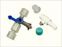

Johns ECG adaptor
Figure 1: Johns ECG adaptor
➧ A nerve stimulator is connected to the epidural catheter via an adapter (Johans ECG adapter, Arrow International, Inc., Reading, USA), (Figure 1). 

➧ The epidural catheter and ECG adapter are primed with sterile normal saline. 

➧ The anode lead of the nerve stimulator is connected to an electrode over the upper or lower extremity as a grounding site. 

➧ The cathode lead of the stimulator is connected to the metal hub of the adapter. 

➧ The nerve stimulator is set at a frequency of 1 Hz with a pulse width of 200 msec. 

➧ Electrical stimulation (1-10 mA) with a segmental motor response (truncal or extremities movement) indicates that the catheter is in the epidural space. 

➧ No motor response indicates that it is not. 

➧ Since it is possible to obtain vigorous and uncomfortable twitches with an excessive current, the current output must be carefully increased from zero and stopped once the motor activity is visible. 

➧ Thus, the stimulator used in the test must be sensitive enough to allow a gradual increase of current output from zero up to at least 10 mA. 

➧ Because a motor response will be elicited at a very low current (< 1 mA) in the case of subarachnoid or subdural placement, the current output must be carefully increased in small increments (0.1 mA) between zero and 2 mA.

2-Contrast Fluoroscopy:

➧ Injection of 3 ml contrast medium via the epidural catheter then radiography of spinal cord is done. 

➧ If the epidural catheter is in subdural space, the lateral view will show a contrast medium spreading cephalad over many segments, along the dorsal part of the spinal cord (Figure 2).
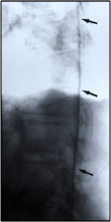
Contrast Fluroscopy, Lateral view
Figure 2: Contrast Fluoroscopy, Lateral view