Educational Blog about Anesthesia, Intensive care and Pain management

Awareness during Anesthesia

Awareness during Anesthesia

Awareness during Anesthesia


Groups at risk for awareness:

Awareness is due to too light plane of anesthesia. It is more likely where muscle relaxants are used. They may be due to:

1-Inadequate anesthetic dose:

-Emergency surgery

-Hypotensive anesthesia

-Cardiac surgery

-Inability to monitor dose in TIVA

-Obstetric surgery

-Normal variability in MAC

2-Resistance:

-Hypermetabolic state

-Smokers

-Obesity

-Alcoholic

3-Equipment malfunction:

-Breathing circuits

-TIVA pump malfunction or line disconnection

-Vaporizers

Prevention:

-Recognize patients at risk

-Anesthesia machine pre-use check and knowing its mechanics & mechanisms

-Periodically check vaporizer level & provide adequate MAC levels

-Benzodiazepines at induction

-Use muscle relaxants ONLY when necessary

-Use of “BIS” monitoring in high-risk groups

-Be aware that some drugs may mask signs of awareness (e.g., β-blockers mask tachycardia)

How to avoid ALI after Thoracic Surgery

How to avoid Acute Lung Injury (ALI) after Thoracic Surgery

-Fortunately, Acute Lung Injury (ALI) occurs infrequently, with an incidence of 2.5 % of all lung resections combined, and an incidence of 8% after pneumonectomy. However, when it occurs, ALI is associated with a risk of mortality or major morbidity of about 40%.

OLV


Causes of ALI:

I. Ventilated Lung:

1-Hyperoxia: (Oxygen toxicity, Reactive oxygen species)

2-Hyperperfusion: (Endothelial damage, Increased pulmonary vascular pressure)

3-Ventilatory Stress: (Volutrauma, Barotrauma, Atelectrauma)

II. Collapsed Lung:

1-OLV: (Ischemia/Reperfusion, Reexpansion, Cytokine release, Altered redox status)

2-Surgery: (Manipulation trauma, Lymphatic disruption)

III. Systemic:

-Cytokine release, Reactive oxygen species, Complement activation, Overhydration, Chemotherapy/Radiotherapy.

Prophylaxis against ALI:

A) Protective Lung Ventilation Strategies:

I. Lower Tidal Volumes (6–8 mL/kg):

-The use of lower tidal volumes may lead to lung derecruitment, atelectasis, and hypoxemia. Lung derecruitment may be avoided by the application of external PEEP and frequent recruitment maneuvers.

II. PEEP (5–10 cm H2O):

-Although PEEP may prevent alveolar collapse and development of atelectasis, it may cause a decrease in PaO2 due to diversion of blood flow away from the dependent, ventilated lung and an increase in the total shunt.

-Thus, PEEP must be customized to the underlying disease of each patient, and a new application of PEEP will rarely be the appropriate way to treat hypoxemia that occurs immediately after the onset of one-lung ventilation.

-Patients with obstructive pathology may develop intrinsic PEEP. In these patients, the application of external PEEP may lead to unpredictable levels of total PEEP.

III. Lower FiO2 (50-80%):

-Although the management of one-lung ventilation has long included the use of 100% oxygen, evidence of oxygen toxicity has accumulated both experimentally and clinically.

-Clinicians recommend titrating FiO2 to maintain the O2 saturation >90%, especially in patients who have undergone adjuvant therapy and are at risk of developing ALI.

IV. Lower Ventilatory Pressures:

-Plateau pressure <25 cm H2O; and peak airway pressure <35 cm H2O, through the use of pressure-controlled ventilation, may diminish the risk of barotrauma.

-The flow pattern results in a more homogenous distribution of the tidal volume and improved dead space ventilation.

V. Permissive Hypercapnia:

-Periodic ABG analysis is helpful to ensure adequate ventilation. End-tidal CO2 measurement may not be reliable due to increased dead space and an unpredictable gradient between the arterial and end-tidal CO2 partial pressure.

VI. At the end of the procedure:

-The operative lung is inflated gradually to a peak inspiratory pressure of less than 30 cm H2O to prevent disruption of the staple line.

-During reinflation of the operative lung, it may be helpful to clamp the lumen of the dependent lung, to limit over-distension.

B) IV Fluids:

-Restrict IV fluids in pulmonary resection to avoid lower lung syndrome (Gravity-dependent transudation of fluid).

What is the meaning of Research?


The word “research” originated from the old French word “researcher” meaning to search and search again.

It literally implies repeating a search for something and implicitly assumes that the earlier search was not exhaustive and complete in the sense that there is still scope for improvement.

Research in common parlance refers to a search for knowledge.

It may be defined as a scientific and systematic search for pertinent information on a specific topic/area. In fact, research is an art of scientific investigation.

The Advanced Learner’s Dictionary of Current English lays down the meaning of research as “a careful investigation or inquiry, especially through search for new facts in any branch of knowledge”. Redman and Mory define research as “a systematized effort to gain new knowledge”. Some people consider research as a movement, a movement from known to unknown. It is actually a voyage of discovery.

Research is a scientific approach to answering a research question, solving a problem, or generating new knowledge through a systematic and orderly collection, organization, and analysis of information with the ultimate goal of making valuable research in decision-making.

Systematic research in any field of inquiry involves three basic operations:

1. Data collection: It refers to observing, measuring, and recording information.

2. Data Analysis: This refers to arranging and organizing the collected data so that we may be able to find out what their significance is and generalize about them.

3. Report writing: It is an inseparable part and a final outcome of a research study. Its purpose is to convey the information contained in it to the readers or audience.

Steps of Systematic Research


Systematic Research follows certain steps that are logical and in order.

These steps are:

1- Understanding the nature of the problem to be studied and identifying the related area of knowledge.

2- Reviewing literature to understand how others have approached or dealt with the problem.

3- Collecting data organized and controlled to arrive at valid decisions.

4- Analyzing data appropriate to the problem.

5- Drawing conclusions and making generalizations.

Characteristics of Research:

Research is a process through which we attempt to systematically and with the support of data to answer a question, resolve a problem, or gain a greater understanding of a phenomenon.

This process has eight distinct characteristics:

Research…

1. Originates with a question or problem.

2. Requires a clear articulation of a goal.

3. Follow a specific plan of procedure.

4. Usually divides the principal problem into more manageable sub-problems.

5. Is guided by the specific research problem, question, or hypothesis.

6. Accepts certain critical assumptions.

7. Requires the collection and interpretation of data in attempting to resolve the problem that

initiated the research.

8. Is by its nature, cyclical; or more exactly, helical.

Ultrasound Artifacts

Ultrasound Artifacts


Ultrasound Artifacts


1-Reverberation artifact:

➧ The processing unit in the ultrasound machine assumes echoes return directly to the processor from the point of reflection. 

➧ Depth is calculated as D = V × T, where V is the speed of sound in biological tissue and is assumed to be 1,540 m/sec, and T is time. 

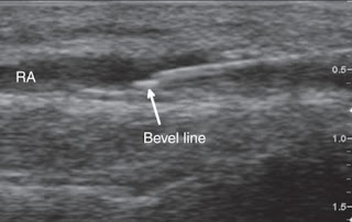
➧ In a reverberation artifact, the ultrasound waves bounce back and forth between two interfaces (the lumen of the needle) before returning to the transducer. 

➧ Since velocity is assumed to be constant at 1,540 m/sec by the processor, the delay in the return of these echoes is interpreted as another structure deep into the needle and hence the multiple hyperechoic lines beneath the block needle (Figure 1). 

Ultrasound Artifacts
Figure 1: Reverberation artifact


2-Mirror artifact:

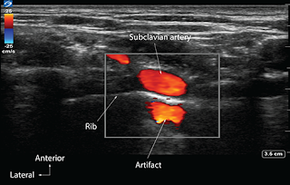
➧ A mirror artifact is a type of reverberation artifact. 

➧ The ultrasound waves bounce back and forth in the lumen of a large vessel (subclavian artery). 

➧ The delay in the time of returning waves to the processor is interpreted by the machine as another vessel distal to the actual vessel (Figure 2). 

Ultrasound Artifacts
Figure 2: Mirror artifact


3-Bayonet artifact:

➧ The processor assumes that the ultrasound waves travel at 1,540 m/sec through biological tissue. However, we know that there are slight differences in the speed of ultrasound through different biological tissues. 

➧ The delay in the return of echoes from tissue that has a slower transmission speed, coupled with the processor’s assumption that the speed of ultrasound is constant, causes the processor to interpret these later returning echoes from the tip of the needle traveling in tissue with slower transmission speed as being from a deeper structure and thus giving a bayoneted appearance. 

➧ If the tip is traveling through tissue that has a faster transmission speed, then the bayoneted portion will appear closer to the transducer (Figure 3). 

Ultrasound Artifacts
Figure 3: Bayonet artifact


4-Acoustic Enhancement artifact:

➧ Acoustic enhancement artifacts occur distal to areas where ultrasound waves have traveled through a medium that is a weak attenuator, such as a large blood vessel. 

➧ Enhancement artifacts are typically seen distal to the femoral and the axillary artery (Figure 4). 

Ultrasound Artifacts
Figure 4: Acoustic enhancement artifact


5-Acoustic shadowing:

➧ Tissues with high attenuation coefficients, such as bone, do not allow the passage of ultrasound waves. 

➧ Therefore any structure lying behind tissue with a high attenuation coefficient cannot be imaged and will be seen as an anechoic region. (Figure 5). 

Ultrasound Artifacts
Figure 5: Acoustic shadowing artifact


6-Absent blood flow:

➧The Color-Flow Doppler may not detect blood flow when the ultrasound probe is perpendicular to the direction of blood flow (Figure 6). 

➧ A small tilt of the probe away from the perpendicular should visualize the blood flow (Figure 7, Figure 8). 

➧ Alternatively, for deep vascular structures, signals may be lost due to attenuation. 

➧ Increasing gain, while in Doppler Color-Flow mode, will increase the intensity of the returning signals, which may detect blood flow that was not previously detected. 

Ultrasound Artifacts
Figure 6: Radial a. Absent blood flow artifact
Ultrasound Artifacts
Figure 7: Radial a. Probe tilted away from the direction of blood flow
Ultrasound Artifacts
Figure 8: Radial a. Probe tilted towards the direction of blood flow

Laryngeal Mask Airway

Laryngeal Mask Airway (LMA)

1-The LMA-Classic™:


-It is a reusable LMA™ airway for general anesthesia.
-The LMA-Classic™ is available in eight sizes: (1, 1½, 2, 2½, 3, 4, 5, and 6).

Advantages:

-A safe and effective alternative to the endotracheal tube and the facemask.

-Over 100 million uses worldwide.

-Leaves the anesthetist's hands free to attend, to monitoring and record keeping.

-Latex-free and exceptionally well tolerated.

-A reusable device that can be cleaned and steam sterilized up to 40 times before being discarded.

2-LMA-Unique™ (Single-use):


-The LMA-Unique™ is a convenient, single-use LMA™ airway suitable for general anesthesia procedures.

-The LMA-Unique™ is available in sizes: (1, 1½, 2, 2½, 3, 4, and 5).

-The LMA-Unique™ is a disposable, single-use device, made to the same design specifications as the LMA-Classic™.

Advantages:

-Packaged sterile - ready for use.

-Suitable for use on emergency vehicles.

-Suitable where access to sterilization facilities is limited.

-Made of medical-grade PVC.

3-LMA-Flexible™:


-The LMA-Flexible™ is a reinforced LMA™ airway with a flexible airway tube.

-The LMA-Flexible™ is available in sizes: (2, 2½, 3, 4, 5, and 6).

-The LMA-Flexible™ is a re-usable device that can be cleaned and steam sterilized up to 40 times before being discarded.

Advantages:

-Designed for ENT, dental, and head surgery.

-Allows extreme flexion.

-Guaranteed kink and crush-proof.

-Latex-free.

4-LMA Flexible (Single-use):


-The LMA Flexible™ Single Use is ideal for use in ENT, ophthalmic, dental, and other head and neck cases and extends the LMA™ Airway benefits of hemodynamic stability and smoother emergence to more procedures.

5-LMA-ProSeal™:


-The LMA-ProSeal™ is an advanced LMA™ airway suitable for general anesthesia.

-It has a unique double cuff arrangement that provides an exceptionally effective, 'hands-free' airway seal, at low intracuff pressures.

-The LMA-Proseal™ is available in sizes: (1½, 2, 2½, 3, 4, and 5).

-The LMA-Proseal™ is a re-usable device that can be cleaned and steam sterilized up to 40 times before being discarded.

Advantages:

-A new double tube design separates the respiratory and alimentary tracts, providing a safe escape channel for regurgitated fluids in the event of unexpected regurgitation.

-The mask is designed to be a minimally stimulating airway device, whose cuff tip presses against the upper oesophageal sphincter when it is correctly positioned. The sides of the mask face the pyriform fossae and the upper border rests against the base of the tongue.

-Latex-free.

6-LMA Supreme™ (Single-use):


-The first and only single-use laryngeal mask with a built-in drain tube.
-The integrated drain tube is designed to channel fluid and gas safely away from the airway. Several simple and quick tests help verify accurate positioning.
-An improved curve for easy insertion. Subtle refinements in the mask make correct placement easier.

7-LMA Fastrach™ & LMA Fastrach™ ETT:


-The design of the LMA-Fastrach™ facilitates rapid insertion from any position, even if space is limited, and moving the patient is a possible hazard.

-The device is self-positioning with the rigid tube designed to fit the curvature of the palatopharyngeal arch, enabling a firm seal to be achieved.

-The LMA-Fastrach™ is available in sizes: (3, 4, and 5).

-The LMA-Fastrach™ is a reusable device that can be cleaned and steam sterilized up to 40 times before being discarded.

Advantages:

The LMA-Fastrach™ has additional features to those of the LMA-Classic™:

-Designed specifically for the anatomically difficult airway.

-Ideal in emergency situations.

-Can be used as an intubating tool, with no interruption of patient oxygenation.

-Allows insertion in the neutral position, in limited space.

-No need to move the patient.

-No need to insert fingers into the patient's mouth.

8-LMA Fastrach™ & LMA Fastrach™ ETT (Single-use):

9-LMA CTrach™:


-The only difficult airway device that allows ventilation, visualization, and intubation.

-The LMA CTrach™ is designed to increase intubation success rates in difficult airways. The LMA CTrach™ mask enables ventilation during intubation attempts while built-in fiber optics provide a direct view of the larynx and real-time visualization of the ET tube passing through the vocal cords.

-The LMA CTrach™ can be inserted exactly the same as the LMA Fastrach™, however, unlike the LMA Fastrach™, once the airway is secured and the patient is being ventilated, the viewer is switched on, placed in the magnetic connector, and a clear image of the larynx is displayed in real-time. The ET tube can be viewed as it enters the trachea. Once the patient is intubated, the viewer is removed and the mask is removed leaving the ET tube in place.

10-i-gel™:


-The i-gel supraglottic airway device accurately and naturally positions itself over the laryngeal framework to provide a reliable peri-laryngeal seal without the need for an inflatable cuff.

-i-gel is made from a medical-grade thermoplastic elastomer, i-gel has been designed to create a non-inflatable, anatomical seal of the pharyngeal, laryngeal, and peri-laryngeal structures whilst avoiding compression trauma.

-i-gel is currently available in sizes: (1, 1½, 2, 2½, 3, 4, and 5), and is supplied in an innovative, color-coded polypropylene ‘cage pack’.

Advantages:

-No inflatable cuff offers easy, rapid insertion.

-An integral bite block reduces the possibility of airway occlusion.

-A buccal cavity stabilizer aids rapid insertion and eliminates potential rotation.

-Made from a unique, soft, gel-like material to allow easy insertion and reduced trauma.

-Gastric channel designed to improve and enhance patient safety.

-Reduces the possibility of epiglottis downfolding and obstructing the airway.

-Unique packaging protects the i-gel in transit and ensures maintaining of its anatomical shape.

Rheumatoid Arthritis

 Rheumatoid Arthritis:

A common, autoimmune connective tissue disease, primarily involving joints, but with widespread systemic effects. There are hypergamma-globulinemia and rheumatoid factors, which are autoantibodies of IgE, IgA, and IgM classes.


Preoperative abnormalities:

1. Articular problems:

-The joint disease involves inflammation, formation of granulation tissue, fibrosis, joint destruction, and deformity. Any joint may be affected. Those of particular concern to the anesthetist is the cervical, the temporomandibular, and the cricoarytenoid joints.

-Airway obstruction can occur from closely adducted, immobile vocal cords, or from laryngeal amyloidosis. Rheumatoid nodules can affect the larynx.

2. Extra-articular problems: occur in more than 50% of patients.

a) Lungs. May be affected by effusions, nodular lesions, diffuse interstitial fibrosis, or Caplan’s syndrome. This is a form of massive pulmonary fibrosis seen in coal miners with rheumatoid arthritis or positive rheumatoid factor and probably represents an abnormal tissue response to inorganic dust. There may be a restrictive lung defect, with a contribution from reduced chest wall compliance.

b) Kidney. Twenty-five percent of patients eventually die from renal failure. Renal damage may be related to the disease process itself, secondary amyloid disease, or drug treatment.

c) Heart. Is involved in up to 44% of cases. Small pericardial effusions are common but are not usually of clinical significance. Rarely, pericarditis and tamponade may occur, usually in seropositive patients and those with skin nodules. Other problems include endocarditis or left ventricular failure. Occasionally heart valve lesions occur and are of two types; rheumatoid granulomas involving the leaflets and ring, and no granulomatous valvular inflammation with thickening and fibrosis of the leaflets.

d) Blood vessels. A widespread vasculitis can occur. Small arteries and arterioles are often involved, frequently in the presence of relatively disease-free main trunk vessels. Significant ischemia may result, in the actual effects depending on the tissue or organ supplied.

e) Autonomic involvement.

f ) Gastrointestinal. Swallowing problems and dysphagia were found in patients with classical rheumatoid arthritis.

g) Peripheral neuropathy.

3. Chronic anemia, which has been shown to respond to erythropoietin therapy, is common.

Anesthetic problems:

1. Disease of the cervical vertebrae. Cervical involvement, and damage to the cervical spinal cord, have been associated with neck manipulation during anesthesia and sedation. Instability is said to occur in 25% of patients with rheumatoid arthritis. Of these, one-quarter will have no neurological symptoms to alert the physician. The problem of instability is not necessarily confined to those with longstanding diseases.

The commonest lesion is atlantoaxial subluxation, although subaxial subluxations may occur in addition. Destruction of bone, and weakening of the ligaments, allow the odontoid peg to migrate backward and upwards, compressing the spinal cord against the posterior arch of the atlas. Thus, the main danger lies in cervical flexion.

The potential dangers of anesthesia and endoscopy have been emphasized. Flexion of the head and reduction in muscle tone may result in cervical cord damage. Dislocation of the odontoid process and spinal cord damage were discovered in a patient undergoing postoperative IPPV in the ITU. It was not known exactly when this had occurred.

2. Cervical instability below the level of a fusion. Those who have previously undergone occipital cervical fusion may develop cervical instability below the level of the original arthrodesis. Occipital-cervical fusion is thought to generate a greater force at the lower cervical level that in turn stresses the unfused facet joints.

3. Laryngeal problems. A constant pattern of laryngeal and tracheal deviation is reported to occur in some patients, particularly those with proximal migration of the odontoid peg. The larynx is tilted forwards, displaced anteriorly and laterally to the left, and the vocal cords are rotated clockwise. Involvement of the larynx in the rheumatoid process is more common than was previously thought. However, fatal airway obstruction occurred following cervical spine fusion, secondary to massive edema in the meso- and hypopharynx.

4. The laryngeal mask airway should not be relied upon to overcome failed tracheal intubation. It was impossible to insert a laryngeal mask airway into a patient with a grade 4 laryngoscopic view. Subsequent cervical X-rays with the head maximally extended showed that the angle between the oral and pharyngeal axes at the back of the tongue was only 70 degrees, compared with 105 degrees in five normal patients. A simulation of different angles using an aluminium plate showed that at an angle less than 90 degrees, the laryngeal mask airway could not be advanced without kinking at the corner.

5. Sleep apneas. Medullary compression associated with a major atlantoaxial subluxation may result in nocturnal oxygen desaturation.

6. Limitation of mouth opening may occur secondary to arthritis of the temporomandibular joints. This is a particular problem in juvenile rheumatoid arthritis.

7. A pericardial effusion and tamponade can be presented as an acute abdominal emergency in patients with seropositive rheumatoid arthritis.

8. Rheumatoid aortic valve involvement may be more rapidly progressive than aortic valve disease from other causes so that there is little time for compensatory hypertrophy of the ventricle to occur. Acute aortic regurgitation caused sudden cardiac failure in a young woman and required urgent valve replacement.

9. Lung disease can result in reduced pulmonary reserve and hypoxia.

10. An increased sensitivity to anesthetic agents may occur.

Management:

1. Clinical assessment of neck and jaw mobility. The Sharp and Purser test gives some indication of cervical spine instability. The patient should be upright, relaxed, and with the neck flexed. With a finger on the spinous process of the axis, the forehead should be pressed backward with the other hand. Normally there is minimal movement. If subluxation is present, the head moves backward as reduction occurs.

2. A lateral view of the cervical spine in flexion and extension will show the distance between the odontoid peg and the posterior border of the anterior arch of the atlas. If subluxation is present, this distance is greater than 3 mm. Frontal views of the odontoid and entire cervical spine have also been suggested.

3. Cervical X-rays of patients who have previously undergone occipital spinal fusions should be carefully examined for evidence of cervical instability at a lower level.

4. Intubation methods. Cervical instability may be an indication of awake fiberoptic intubation with the application of a collar or Crutchfield tongs, to maintain rigidity during surgery. Since spinal instability is usually in flexion, some authors believe that safe tracheal intubation can be achieved under general anesthesia by careful extension of the head, except in the rare instances of posterior atlantoaxial subluxation when fibreoptic intubation is indicated. Emergency control of the airway has been described using a laryngeal mask airway in a patient who developed acute pulmonary edema following occipital-cervical fusion.

5. Deviation of the larynx may make fibreoptic laryngoscopy more difficult in some patients. Examination of the orientation of the larynx by indirect laryngoscopy at preoperative assessment may be helpful. If there is cricoarytenoid involvement, care should be taken with the choice of tracheal tube size and tube insertion. Cricoarytenoid arthritis may occasionally necessitate permanent tracheostomy.

6. Although the use of the laryngeal mask airway is increasingly common, as mentioned above, it cannot always be relied on in patients with severe flexion deformities of the neck.

7. Assessment of pulmonary function and reserve.

8. Examination for other significant complications, such as valvular disease, or pericardial effusion.

9. Extreme caution should be observed if epidural or caudal anesthesia is to be undertaken in patients in whom intubation difficulties are anticipated. Even after a test dose to exclude an accidental spinal, or vascular penetration, the block should only be established very gradually.

10. The use of cervical epidural analgesia for the treatment of digital vasculitis has been reported.

Anesthetic Considerations for Patients with Liver disease

Anesthetic Considerations for Patients with Liver Disease


Preoperative:

1. Assess the Degree of hepatic impairment, Severity, and Hepatic reserve by the Child-Turcotte-Pugh scoring system.

2. AVOID: Premedication, IM injections, Contact with blood or body fluids, unnecessary esophageal instrumentation.

Regional Anesthesia:

-Regional anesthesia might be used when possible in patients with advanced liver disease.

-Coagulopathy (PT & INR) should be considered a contraindication to some types of regional anesthesia.

-AVOID Epidural a. (Large amounts of amide LAs).

IV Anesthetics:

-Propofol, Ketamine (in hypotensive patients).

Opioids:

-Opioids can also be used successfully in patients with the hepatic disease despite certain pharmacological consequences (decreased clearance and prolonged half-life).

-Fentanyl is considered the opioid of choice because it does not decrease hepatic oxygen and blood supply nor does it prevent increases in hepatic oxygen requirements when used in relatively moderate doses.

-AVOID Morphine (Active metabolite, Prolonged action).

Changed Pharmacokinetics:

-The half-life of lidocaine in patients with liver disease may be increased by more than 300%, for benzodiazepines by more than 100%, etc.

-For drugs binding to albumin, the volume of distribution is decreased and therefore the drug dosage should be decreased (e.g. sodium pentothal).

Muscle Relaxants:

-Suxamethonium → Prolonged action, Atracurium, Cisatracurium (of choice).

-AVOID Pancuronium, Vecuronium (Hepatic metabolism).

-The volume of distribution of many drugs can be substantially increased (for different reasons, including an increase in gamma globulin and edema), dictating a necessity to increase the first effective dose of the drug.

-However, owing to a decrease in hepatic blood flow and hepatic metabolic and excretory functions, as well as impaired renal function, the clearance of such a drug is decreased, therefore the effect can be prolonged (e.g. pancuronium).

-Atracurium has a theoretical advantage because its metabolism is not dependent on liver function. Therefore, the clearance and elimination half-life of atracurium in patients with impaired hepatic and/or renal function is not different from those with normal hepato-renal function. However, the volumes of distribution are larger, and, accordingly, the distribution half-lives are shorter in patients with severe hepato-renal dysfunction compared with normal individuals.

-Titration of any relaxant according to the transcutaneous nerve stimulation monitoring is beneficial because the degree of hepatic dysfunction affects the degree of pharmacokinetic disorders.

Inhalational Anesthetics:

-Halothane should be avoided because it leads to the most prominent decrease in hepatic blood flow and oxygen supply and postoperative hepatic dysfunction. In addition, immunologically mediated severe postoperative halothane hepatitis may follow halothane anesthesia.

-Isoflurane is a better choice if an inhalational technique is selected.

-More recently introduced volatile anesthetics, sevoflurane, and desflurane, each of them, can be used safely in patients with liver disease, as they preserve hepatic blood flow.

-Nitrous oxide has been used in patients with advanced hepatic disease for many years, and so far has not been incriminated in increased anesthesia-related hepatic postoperative complications. However, a well-known sympathomimetic effect of nitrous oxide and some possibilities of jeopardizing oxygenation render the routine use of nitrous oxide in patients with advanced liver disease undesirable. It is important to remember that long surgical operations under anesthesia with nitrous oxide might result in the accumulation of nitrous oxide in the intestinal lumen with subsequent intestinal distension.

Others:

-Renal function must be maintained by administering proper fluid load (volume and content); (avoid Na+ overload, use glucose-containing solutions for hypoglycemia, albumin 5% is the preferred colloid), and diuretics if needed.

-The parameters of controlled ventilation should be carefully selected to avoid an unnecessary increase in intrathoracic pressure which may impede venous return thereby decreasing cardiac output.

-Monitoring the coagulation state during surgery can be important. The treatment should be based on the results of hematologic monitoring and may include administration of platelets, fresh frozen plasma, cryoprecipitate, and sometimes tranexamic acid.

IV Fluids

IV Fluids

A) Crystalloid solutions (< 30 000 Dalton):

1-Hypotonic solutions:

- D5W, ½ NS, D5 ¼ NS, D5 ½ NS.

2-Isotonic solutions:

- NS, Ringer’s, Lactated Ringer’s (Hartmann’s), Ringer’s acetate.

3-Hypertonic solutions:

- NaCl (3%, 7.5%), Mannitol (10%, 20%), NaHCO3 (5%, 7.5%, 8.4%).

B) Colloid solutions (> 30 000 Dalton):

1-Blood derived:

- Human albumin (5%, 25%).

- Plasma protein fraction 5%.


2-Synthetic:

a) Dextrose Starches:

- They are D-glucose polymers linked by alpha bonds into linear macromolecules prepared from sucrose.

- Their half-life is 12 hours.

- 6% Dextran-70 (Macrodex) MW = 70 000 Dalton

- 10% Dextran-40 (Rheomacrodex) MW = 40 000 Dalton

- Dextran-1 (Promit).

- Dose: 20 ml/kg/d., Max.: 1.5 L/d.


Disadvantages:

- Interfere with blood typing.

- Antiplatelet effect.

- Antigenic effect with anaphylactoid and anaphylactic reactions.

- Affects renal functions and can be associated with renal failure.

b) Gelatins:

- Their M.W. 30 000-35 000 Dalton.

- Their half-life is 4-5 hours.

- Succinylated Gelatin (Gelofusine 3.5%, 4%).

- Polygelline (Hemaccel 3.5%, Hemagel 4%).

- Dose: 20 ml/kg/d., Max.: 1.5 L/d.


Advantages:

- Does not interfere with blood grouping.

- Does not affect renal function.

Disadvantages:

- Histamine-mediated allergic reaction.

c) Hydroxy-Ethyl Starch (HES) (Etherified Starch):

Synthetic colloids resemble glycogen structurally, synthesized from amylopectin.

- Heta-starch (Hexa-starch) (e.g. HAES-Steril, Hespan): M.W. 450 000 Dalton, half-life 48 hours.

- Hextend: M.W. is higher, and the half-life is longer.

- Penta-starch: M.W. 200 000, half-life less than 24 hours.

- Tetra-starch: (e.g. Tetraspan, Voluven): M.W. 130 000, half-life less than 6 hours.

- Dose: 20-50 ml/kg/d.


Advantages:

- Do not affect the coagulation study.

- Not antigenic but can cause anaphylactoid reactions.

- Less expensive than human albumin.