Educational Blog about Anesthesia, Intensive care and Pain management

Cell (Blood) Salvage

Cell (Blood) Salvage

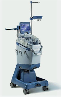
Cell Saver Machine


Definition:

-Cell (blood) salvage is a process in which a patient’s own (lost) blood is collected, processed, and transfused back (‘Autologous’ blood transfusion), which is done by a cell saver machine.

Principle:

1-Collection of blood: blood is suctioned from the operative field, and then heparinized saline is added, filtered, and centrifuged to separate RBCs which are then washed.

2-Washing of RBCs: across a semi-permeable membrane to filter out free Hb, plasma, WBCs, and platelets.

3-Re-transfusion: Washed RBCs are then suspended in saline (to achieve Hct of 60-70%) and transfused within 6 hours.

Advantages:

-Reduce or eliminate the need for ‘allogenic’ transfusion.

-‘Allogenic’ blood transfusion has been associated with an increased risk of postoperative infection, acute lung injury, perioperative MI, low CO HF, and tumor recurrence.

Indications:

-Anticipated blood loss >1L or >20% of estimated blood volume

-Patients with low Hb, multiple RBCs alloantibodies, rare blood group, and patient refusal of ‘allogenic’ blood transfusion

-Obstetrics: Controversial due to potential risk of amniotic fluid embolism. However; cell salvage with a Leucocyte-depletion filter (LDF) is considered safe

-Orthopedics: Reduce ‘allogenic’ transfusion & postoperative infection in arthroplasty

-Cardiac surgery: Leucocyte-depletion filter (LDF) use, reduce micro-emboli & lipid load of cell salvaged blood with an improvement of postoperative lung function.

-Vascular surgery

-Liver transplantation

-Jehovah’s Witness

Contraindications:

-Malignancy: due to risk of tumor dissemination

-Wound contamination: due to risk of systemic spread

-Old hemolyzed blood

-Use of collagen or hemostatic materials

-Obstetric surgery: due to risk of amniotic fluid embolism

-Ascites

Complications:

-Non-immune hemolysis: due to centrifugation

-Coagulopathy: due to large volumes of transfusion

-Citrate overdosage

-Air embolism

-Febrile non-hemolytic transfusion reaction

-Contamination: due to incomplete washing leading to contamination with drugs, activated leukocytes, cytokines, and microaggregates.

Dexmedetomidine

Dexmedetomidine

Mechanism of Action:

-It is an imidazole derivative and is a specific alpha-2 adrenoceptor agonist that acts via post-synaptic alpha-2 receptors primarily in the locus ceruleus to increase conductance through K+ channels.

Dexmedetomidine


Uses and Dose:

-Its main actions are sedation, anxiolysis, and analgesia

-It is a clear, colorless isotonic solution containing 100 g/ml of dexmedetomidine base and 9 mg/ml of sodium chloride in water. The solution is preservative-free and contains no additives.

-Dexmedetomidine can be administered intravenously, intramuscularly, and transdermally.

1. ICU Sedation:

-Used for sedation of initially intubated and mechanically ventilated patients in ICU.

-Loading: 1 mcg/kg IV over 10 minutes; loading dose may not be required for adults converted from other sedative therapy.

-Maintenance 0.2-0.7 mcg/kg/h. continuous IV infusion; not to exceed 24 h.

-The duration of use should not exceed 24 hours.

-Dexmedetomidine has been infused in mechanically ventilated patients before, during, and after extubation; it is not necessary to discontinue dexmedetomidine before extubation.

2. Procedural Sedation:

Indicated for sedation of non-intubated patients before and/or during surgical and other procedures.

Loading: 1 mcg/kg IV over 10 minutes.

Maintenance 0.6 mcg/kg/h. IV titrate to effect (usually 0.2-1 mcg/kg/h.).

3. Awake Fiberoptic Intubation:

Loading: 1 mcg/kg IV over 10 minutes.

Maintenance 0.7 mcg/kg/hr IV until endotracheal tube secured.

Dosage Modifications:

-Dose reduction may be required if co-administered with other concomitant anesthetics, sedatives, hypnotics, or opioids.

-Consider dose reduction in patients with hepatic impairment or aged ≥ 65 y.; clearance decreases with increasing severity of hepatic impairment.

-Renal impairment: No dosage adjustment required.

Pharmacokinetics

Distribution: It is 94% protein-bound in the plasma; the volume of distribution is 1.33 l/kg. The distribution half-life is 6 minutes.

Metabolism: The drug undergoes extensive hepatic metabolism to methyl and glucuronide conjugates.

Excretion: 95% of the metabolites are excreted in the urine. The elimination half-life is 2 hours, and the clearance is 39 l/hour.

Pharmacodynamics:

1. Cardiovascular System: It causes a predictable decrease in the mean arterial pressure and heart rate.

2. Respiratory System: It causes a slight increase in PaCO2 and a decrease in minute ventilation, with minimal change in the respiratory rate—these effects are not clinically significant.

3. Central Nervous System: The drug is sedative and anxiolytic—ventilated patients remain easily arousable and cooperative during treatment. Reversible memory impairment is an additional feature.

4. Metabolic / Other: It causes a decrease in plasma epinephrine and norepinephrine concentrations. It does not impair adrenal steroidogenesis when used in the short term.

Side Effects:

Hypotension, bradycardia, nausea, and a dry mouth are the most commonly reported side effects of the drug.

Anesthesia for Electroconvulsive Therapy

Anesthesia for Electroconvulsive Therapy

Anesthesia for Electroconvulsive Therapy


Principle:

-The exact mechanism of Electroconvulsive Therapy (ECT) is unknown. Electrical stimuli (electroconvulsive shock) are usually administered until a therapeutic generalized seizure is induced (30–60 sec. in duration).

-A good therapeutic effect is generally not achieved until a total of 400–700 seizures have been induced, in several sessions, over 2-3 weeks. Progressive short memory loss often occurs with an increasing number of treatments.

Physiological Effects:

-Seizure activity is characteristically associated with an “initial parasympathetic” discharge characterized by bradycardia and increased secretions. Marked bradycardia (<30 beats/min.) and even transient asystole (up to 6s) are occasionally seen.

-This is followed by “sustained sympathetic” discharge. Hypertension and tachycardia that follow are typically sustained for several minutes.

-Transient autonomic imbalance can produce arrhythmias and T-wave abnormalities on the ECG. Cerebral blood flow and ICP, intragastric pressure, and intraocular pressure all transiently increase.

Contraindications:

• Recent MI (<3 months)

• Recent stroke (usually <1 month)

• Intracranial mass or increased ICP from any cause

• More relative contraindications include:

- Angina

- Poorly controlled heart failure

- Significant pulmonary disease

-Bone fractures, Severe osteoporosis

- Pregnancy

- Glaucoma and retinal detachment.

Anesthetic Considerations:

-Amnesia is required only for a brief period (1–5 min) from when the NMB is given to when a therapeutic seizure has been successfully induced. The seizure itself usually results in a brief period of anterograde amnesia, somnolence, and often confusion. Consequently, only a short-acting induction agent is necessary.

-Increases in seizure threshold are often observed with each subsequent ECT.

-Most induction agents (Barbiturates, Benzodiazepines, and Propofol) have anticonvulsant properties, small doses must be used. The seizure threshold is increased and seizure duration is decreased by all of these agents.

--Sodium pentothal (2–4 mg/kg) was the first induction agent used, it raises the seizure threshold and decreases its duration.

--Methohexital (0.5-1.0 mg/kg): has been the induction agent of choice (gold standard) because it has very little effect on seizure duration and has a rapid onset and recovery profile. Unfortunately, methohexital is no longer available.

--Benzodiazepines: raise the seizure threshold and decrease its duration.

--Propofol (1–1.5 mg/kg): but higher doses reduce seizure duration.

--Etomidate (0.15-0.6 mg/kg): lacks anticonvulsant properties, increases seizure duration, and prolongs recovery.

--Ketamine (1.5-2 mg/kg): lacks anticonvulsant properties, and increases seizure duration, but is generally not used because it also increases the incidence of delayed awakening, nausea, and ataxia and is also associated with hallucinations during emergence.

-Short-acting opioids: are not given alone because they do not consistently produce amnesia.

-Sevoflurane (5%–8% for induction, followed by 1–2 MAC): is the only inhalational agent in widespread use for induction in ECT, with comparable effects to intravenous (IV) agents. It is preferred for patients not cooperative with IV access. It has the advantage of attenuating uterine contractions following ECT and is used in the third trimester of pregnancy.

-Induction agents in the descending order of seizure duration after their use are:

[Etomidate > Ketamine > Methohexital > Sevoflurane > Thiopental > Propofol]

-Induction agents in descending order of seizure threshold reducing property are:

[Etomidate > Ketamine > Methohexital > Thiopental > Propofol]

-Neuromuscular blockade: required from the time of electrical stimulation until the end of the seizure. A short-acting agent, such as succinylcholine (0.25–0.5 mg/kg), is most often selected.

-Ventilation: Controlled “mask ventilation” (with a backup plan of LMA if concerned about effective ventilation), is required until spontaneous respirations resume. As ECT is usually administered 3-times a week, repeated intubations may lead to airway trauma and edema. Hyperventilation can increase seizure duration and is routinely employed in some centers.

Monitoring:

-Routine monitoring should be as with the use of any other general anesthetic.

-Seizure activity is monitored by an unprocessed EEG. It can also be monitored in an isolated limb: a tourniquet is inflated around one arm before injection of succinylcholine, preventing entry of the NMB and allowing observation of convulsive motor activity in that arm.

Precautions:

-Rubber bite block: to avoid dental, tongue, and lips injury.

-Exaggerated parasympathetic effects: should be treated with atropine. In fact, premedication with glycopyrrolate is desirable both to prevent the profuse secretions associated with seizures and to attenuate bradycardia.

-Sympathetic manifestations: Nitroglycerin, Nifedipine, and α- and β-adrenergic blockers have all been employed successfully for control. High doses of β-adrenergic blockers (Esmolol, 200 mg), however, are reported to decrease seizure duration.

-Patients with pacemakers: may safely undergo ECT treatments, but a method to convert the pacemaker to a fixed mode, if necessary should be readily available.

Post-Tonsillectomy Bleeding

Post-Tonsillectomy Bleeding

Post-Tonsillectomy Bleeding


Incidence and Initial Management:

-The incidence of post-tonsillectomy bleeding ranges from 2% to 4%. It is more common in teenagers and young adults than in small children. The vast majority of postoperative bleeding occurs between days 5 and 10 when the eschar separates from the tonsil beds. In rare cases, bleeding may occur in the immediate postoperative period.

-Initial management is tailored to the degree of bleeding. The clot in the tonsillar bed should be suctioned, as spontaneous hemostasis is rare with the clot in place.

-Minimal bleeding may resolve with an ice water gargle or hydrogen peroxide gargle, and older children may tolerate the application of a cautery stick with silver nitrate while awake.

-Administration of antifibrinolytics as tranexamic acid.

-Significant bleeding in a young child, however, typically involves a return to the operating room for a thorough examination of the pharynx and electrocautery hemostasis.

Preoperative Assessment and Optimization:

1-Airway Assessment:

-Although it may be difficult to assess the airway in-depth in an agitated child, observation of the external anatomy and information about airway management from the prior anesthetic should provide sufficient information.

-Even though airway management was uncomplicated previously, it may be more difficult at this time because of postoperative edema and blood-obscuring visualization of the larynx.

2-Intravascular Volume Status:

-It is possible to underestimate the degree of blood loss, since much of it may have been swallowed. Heart rate, blood pressure, and, if possible, orthostatic testing will provide information regarding volume status and guide volume resuscitation.

-Assessing for the presence of tears, moist mucus membranes, skin turgor, and urine output will be helpful as well.

-Adequate IV access, with a large-bore intravenous catheter, should be obtained, if not already present.

-Intraosseous access should be entertained in the hypovolemic, hypotensive patient if IV access cannot be obtained in a timely fashion.

-Volume resuscitation should be initiated with non-glucose-containing isotonic fluids. If the patient is hypotensive, 20 mL/kg boluses of isotonic fluid should be administered until the blood pressure normalizes.

-Transfuse red blood cells if hemodynamically unstable and hematocrit is low.

-Once the bleeding has been controlled and the patient is normovolemic, maintenance fluids should be continued.

3-Laboratory Tests:

-Hematocrit, platelet count, electrolytes, and coagulation studies.

-A specimen should also be sent for serotyping and cross match, as transfusion is a very real possibility.

-Surgery should not be delayed waiting for test results, since bleeding will continue without surgical intervention

Intraoperative Management:

-Management of the airway is of major concern, particularly the ability to visualize the larynx in the presence of ongoing bleeding.

-In preparation for induction, the following should be prepared: multiple laryngoscope blades, a styletted cuffed ETT, and large bore suction.

-The otolaryngologist should be present and tracheostomy equipment immediately available should a surgical airway become necessary.

-The patient is considered to have a full stomach, even if they have not recently eaten, because of swallowed blood.

-A rapid sequence induction is usually performed. However, in the situation where there is significant concern about the airway, a smooth mask inhalation induction with cricoid pressure can be performed with the patient in the right lateral decubitus position with the head down (tonsillectomy position) with suction immediately available. The tonsillectomy position minimizes aspiration risk by promoting the pooling of blood in the oropharynx.

-The choice of IV induction agent will depend on the volume status. Ketamine or etomidate may be used if there is ongoing concern about volume status and hemodynamic stability. Alternatively, propofol may be used but in a decreased dose.

-Muscle relaxation can be achieved with either succinylcholine or rocuronium. However, the duration of action of rocuronium at the dose recommended for rapid sequence induction (1.2 mg/kg) will likely exceed the length of the procedure.

-Volume status should be continually assessed in the face of ongoing bleeding and managed appropriately with isotonic fluids.

-The hematocrit, degree of hemodynamic stability, and status of hemostasis will dictate the need for a blood transfusion.

-Replacement of coagulation factors is rarely necessary. However, if a previously undiagnosed coagulopathy is discovered, the appropriate therapy should be initiated and a hematology consult obtained.

Postoperative Management:

-Patients should be placed in the lateral decubitus position with the head down with supplemental oxygen for transport to the PACU.

-Analgesic and antiemetic medications should be ordered.

Multiple Sclerosis

 Multiple Sclerosis

Multiple Sclerosis


-Multiple sclerosis (MS) is the most common demyelinating neurological disease.

-The myelin surrounding an axon may develop normally and be lost later, but leaving the axon preserved. Alternatively, there may be some defect in the original formation of myelin as a result of an error in metabolism.

-Multiple sclerosis is thought to be autoimmune in nature. Susceptibility to MS may be genetically determined. Viral and immune factors are possibly involved.

-It is characterized by a combination of inflammation, demyelination, and axonal damage in the CNS. Disruption of the blood-brain barrier is an early event. Plaques of demyelination are scattered throughout the nervous system, usually in the optic nerve, brainstem, and spinal cord. The peripheral nerves are not involved.

Preoperative Findings:

1. The commonest presenting symptoms, in order of frequency, are limb weakness, visual disturbances, paresthesia, and incoordination. Legs are more commonly involved before the arms, with signs of spasticity and hyperreflexia. Urinary symptoms may occur.

2. Progression, with remissions and relapses, is very variable. Infection, trauma, and stress may be associated with relapses. A small increase in body temperature can cause a definite deterioration in neural function. The third trimester of pregnancy is associated with a 70% decrease in relapse rate, but this is followed by an increase of about 70% in the first 3 months postpartum. This may impair the ability of a mother to care for her baby.

3. Pain may be a prominent feature, occurring in 45% of patients.

4. Mild dementia and dysarthria may appear as the disease progresses.

5. In advanced disease, and sometimes earlier during acute relapses, respiratory complications may occur secondary to a variety of causes; they were, in decreasing order of importance, respiratory muscle weakness, bulbar weakness, and central control of breathing.

6. MRI now plays an important part in the diagnosis, and abnormalities in the white matter can be seen in 99% of cases. Gadolinium enhancement seems to reflect areas of inflammation where the blood-brain barrier has broken down.

7. Patients are treated with; baclofen, gabapentin, or beta interferon.

Anesthetic Problems:

1. Both experimentally and clinically, an increase in body temperature has been shown to cause a deterioration in nerve conduction and neurological signs.

2. Spinal anesthesia is associated with an increased incidence of neurological complications.

3. Epidural anesthesia in pregnant women with MS showed that there was no difference between those who had been given an epidural and those who had not. Temporary neurological deficits have, however, been reported. It was postulated that neurotoxicity might have resulted from the diffusion of the local anesthetic into the dural space. However, it has been suggested that concentrations of bupivacaine not greater than 0.25% should be used since postpartum relapse has been reported with those above this.

4. Local anesthesia did not significantly increase the relapse rate. However, early disruption of the blood-brain barrier in MS means that local anesthetics can cross more readily, and toxicity is more likely to occur.

5. Neuromuscular blockers. Resistance to atracurium, in association with an abnormally high concentration of skeletal muscle acetylcholine receptors, has been reported in a patient with MS and spastic paraparesis.

6. There is an increased incidence of epilepsy in MS patients.

Anesthetic Management:

1. Elective surgery should not be undertaken in the presence of fever.

2. Spinal anesthesia should probably be avoided. If a regional block is required, epidural anesthesia is preferable.

3. The maximum dose of local anesthetic should be reduced below that normally recommended. Techniques that require large doses should be avoided.

4. It was suggested that IV gamma globulin immediately after delivery protects patients from relapse in the first 6 months postpartum.

5. Patients may require treatment for pain and spasticity.

Complications of Total Parenteral Nutrition (TPN)

Complications of Total Parenteral Nutrition (TPN)

Complications of Total Parenteral Nutrition (TPN)


I. Catheter-Related Complications:

-The hyperosmolarity of the dextrose and amino acid solutions requires infusion through large veins or central venous lines.

1-Misdirected Catheter: e.g., with subclavian vein cannulations – (mostly on the right) 10% resulted in misplacement of the catheter in the internal jugular vein.

2-Infection

3-Hematoma

4-Thrombosis

II. Carbohydrate Complications:

1-Hyperglycemia:

-Hyperglycemia is common during TPN; blood glucose levels >300 mg/dl were recorded in 20% of postoperative patients receiving TPN.

-A standard TPN regimen with 1,800 non-protein calories has ≈350 g of glucose, compared to 230 g in a standard tube feeding regimen.

-Tight glycemic control is not recommended in critically ill patients because of the risk of hypoglycemia, which has more serious consequences than hyperglycemia.

-The current recommendation for hospitalized patients is a target range of 140–180 mg% for blood glucose.

2-Insulin:

-If insulin therapy is required, regular insulin is preferred for critically ill patients, to prevent wide swings in glucose levels, by adding insulin to the TPN solutions.

-One shortcoming of IV insulin infusions is the propensity for insulin to adsorb to the plastic tubing in IV infusion sets. This affects the bioavailability of insulin but can be reduced by priming the IV infusion set with an insulin solution (e.g., 20 mL of saline containing 1 unit/mL of regular insulin). But the priming procedure must be repeated each time the IV infusion set is changed.

-SC insulin can be used for stable patients. Regimens will vary in each patient, with a combination of intermediate or long-acting insulin with rapid-acting insulin, when needed.

3-Hypophosphatemia:

-The movement of glucose into cells is associated with a similar movement of phosphate into cells, and this provides phosphate for co-factors (e.g., thiamine pyrophosphate) that participate in glucose metabolism. This intracellular shift of phosphate can result in hypophosphatemia.

4-Hypokalemia:

-Glucose movement into cells is also accompanied by an intracellular shift of potassium (which is the basis for the use of glucose and insulin to treat severe hyperkalemia). This effect is usually transient, but continued glucose loading during TPN can lead to persistent hypokalemia.

5-Hypercapnia:

-Excess carbohydrate intake promotes CO2 retention in patients with respiratory insufficiency. This was originally attributed to the high respiratory quotient (VCO2/VO2) associated with carbohydrate metabolism. However, CO2 retention is a consequence of overfeeding, and not overfeeding with carbohydrates.

III. Lipid Complications:

-Overfeeding with lipids may contribute to hepatic steatosis.

-Triggering inflammatory response: The lipid emulsions used in TPN regimens are rich in oxidizable lipids, and the oxidation of infused lipids will trigger an inflammatory response. (Oleic acid, one of the lipids in TPN, is a standard method for producing ARDS in animals), and this might explain why lipid infusions are associated with impaired oxygenation.

IV. Hepatobiliary Complications:

1-Hepatic Steatosis:

-Fat accumulation in the liver (hepatic steatosis) is common in patients receiving long-term TPN and is believed to be the result of chronic overfeeding with carbohydrates and lipids. Although this condition is associated with elevated liver enzymes, it may not be a pathological entity.

2-Cholestasis:

-The absence of lipids in the proximal small bowel prevents cholecystokinin-mediated contraction of the gallbladder. This results in bile stasis and the accumulation of sludge in the gallbladder and can lead to acalculous cholecystitis.

V. Bowel Sepsis:

-The absence of nutritional bulk in the GI tract leads to atrophic changes in the bowel mucosa and impairs bowel-associated immunity, and these changes can lead to the systemic spread of enteric pathogens.

Tetralogy of Fallot

 Tetralogy of Fallot

Tetralogy of Fallot


-Tetralogy of Fallot (TOF) is a congenital cardiac abnormality. The primary defects are pulmonary infundibular stenosis and ventricular septal defect (VSD). The VSD is sufficiently large for the pressure in both ventricles to be equal to that of the aorta. The tetralogy is completed by two secondary features, a variable degree of overriding of the aorta, and right ventricular hypertrophy.

-Dynamic right ventricular outflow obstruction may occur (infundibular spasm), which is accentuated by sympathetic stimulation. The fraction of the right to left-shunt depends primarily upon the relative resistances between the pulmonary (or right ventricular) and systemic outflows.

-If TOF is associated with patent foramen ovale (PFO) or atrial septal defect (ASD), it is a Pentalogy of Fallot.

-The aim of the surgery is to relieve the right ventricular outflow obstruction and to close the VSD. The traditional management by a two-stage repair has been replaced by definitive correction. Recent surgical advances include conduits to connect the RV to the PA and transatrial repair of the VSD.

The problems encountered during anesthesia will depend upon whether or not corrective surgery has been undertaken, and the functional result.

Preoperative Findings:

1. Dyspnea may occur on exertion and is hypoxia-related. Cyanosis and finger clubbing are variable, depending on the degree of pulmonary stenosis and the size of the shunt. Polycythemia (erythrocytosis) is common. There is a pulmonary stenotic murmur, but no murmur from the VSD because of the size of the defect. Squatting is thought to reduce the fraction of the shunt since kinking the large arteries increases systemic vascular resistance. Squatting is commonly seen in children with uncorrected lesions.

2. ECG shows right atrial and right ventricular hypertrophy, right axis deviation, and right bundle branch block.

3. Echocardiography shows right atrial and right ventricular hypertrophy, VSD, PFO or ASD, pulmonary hypertension, and overriding of the aorta.

4. Chest X-ray shows right ventricular hypertrophy and oligemic lungs. In the 2.6–6% of individuals who also have an absent pulmonary valve, aneurysmal dilatation of the pulmonary arteries may cause bronchial compression.

5. Initial surgery may have been undertaken to anastomose a systemic to a pulmonary artery, to improve the pulmonary blood flow, and reduce cyanosis. A definitive procedure is now more commonly undertaken in infancy.

6. In patients who have undergone shunt surgery without a definitive repair, there is chronic hypoxia and polycythemia. These patients have a high mortality, and an increased risk of bacterial endocarditis, thrombotic stroke, emboli, and intracerebral abscess.

7. In adults who have undergone repair there is an increased risk of arrhythmias, conduction defects, and sudden deaths, possibly related to mechanical events, such as ventricular dilatation and stretch, in the proximity of the conduction system. However, new approaches to surgery may result in a lower incidence of such problems in the future.

Anesthetic Problems:

1. In individuals with uncorrected lesions, the right to left shunt, and hence the cyanosis, is increased by a reduction in systemic vascular resistance produced by systemic vasodilatation. This may result from factors such as hypovolemia, drug effects, or pyrexia.

2. Cyanosis is also worsened by an increase in pulmonary vascular resistance or spasm of the right ventricular infundibulum. Right ventricular outflow obstruction is due to increased contractility which is produced by increases in catecholamine output or the administration of drugs with positive inotropic effects. Anxiety, pain, hypercarbia, hypoxia, and acidosis are all precipitating factors. These cyanotic attacks or ‘tet’ spells, which can occur when awake or under anesthesia, may initiate a cycle of increasing hypoxia that can result in cerebral damage or death. Direct intraoperative observations of shunt direction and flow have been made with Doppler color flow imaging using epicardial leads. Patients with severe life-threatening hypoxemic spells, refractory to other treatments, responded to phenylephrine (5 mcg/kg plus an infusion of 0.4–2 mcg/kg/min).

3. Dehydration in the presence of polycythemia and high plasma viscosity may combine to increase the incidence of cerebral thrombosis. Polycythemia may also be associated with coagulation defects.

4. In patients with an absent pulmonary valve, positional airway compromise occurred secondary to bronchial compression of dilated pulmonary arteries.

5. A significant incidence of tracheal anomalies has been found.

6. In adults who have undergone repair, ventricular and atrial arrhythmias are common, particularly during exercise. It has been suggested that patients scheduled for elective surgery should have Holter monitors, or undergo exercise testing, in case antiarrhythmic treatment is needed first.

7. Cyanosed patients with Fallot’s rarely become pregnant. However, adults who have undergone corrective surgery are increasingly present during pregnancy with a favorable outcome.

Anesthetic Management:

1. Antibiotic prophylaxis against bacterial endocarditis.

2. A good premedication (morphine sulphate, or midazolam) to prevent excitement and anxiety.

3. In patients with cyanosis, measures are aimed at reducing the right to left shunt. Specific treatments for cyanotic attacks include:

a) Oxygen 100% to decrease PVR.

b) Pressor agents, such as phenylephrine (5-10 mcg/kg), to increase systemic vascular resistance.

c) Fluids to correct hypovolemia.

d) Propranolol (0.1 mg/kg) or Esmolol (0.5 mg/kg) to decrease outflow tract obstruction (decrease contractility and infundibular spasm).

e) Deepening of light anesthesia to reduce tachycardia associated with catecholamine output.

f) Compression of the femoral artery or the abdominal aorta against the vertebrae, sufficiently firmly to stop the femoral artery pulsations.

4. Techniques to avoid hypoxia and hypercarbia, and minimize vasodilatation and sudden increases in cardiac output. Ketamine, and Morphine sulphate (0.05-0.1 mg/kg), have been used.

5. Hydration is maintained in the perioperative period and, if there is severe polycythemia, venesection may be necessary.

6. Metabolic acidosis should be prevented or treated with sodium bicarbonate (1-2 mmol/kg).

7. In pregnancy, the outcome is satisfactory in patients whose ventricular function is good, and in whom no residual shunt occurs. However, close observation should be undertaken by an experienced team.

8. Venesection for erythrocytosis that is associated with cyanotic congenital heart disease should only take place if there are symptoms of hyperviscosity with a hematocrit >65%, and only provided volume replacement takes place at the same time.

9. Avoid muscle relaxants with histamine release e.g. atracurium, and use cisatracurium or rocuronium.

9. Mechanical ventilation:

-Increase FiO2

-Avoid increased airway pressure (< 15 cmH2O)

-Increase respiratory rate

-Avoid PEEP

Eisenmenger’s Syndrome

 Eisenmenger’s Syndrome

Eisenmenger’s Syndrome


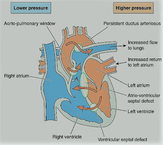
-A rare syndrome of pulmonary hypertension associated with a reversed or bidirectional cardiac shunt, occurring through a large communication between the left and right sides of the heart. The defect may be interventricular, interatrial, or aortopulmonary.

-The development of Eisenmenger’s syndrome, from the initial left to right shunt, is usually a gradual process. Contributory factors to pulmonary hypertension are hypoxia, high pulmonary blood flow, and high left atrial pressure.

-Irreversible structural changes take place in the small vessels, causing pulmonary vascular obstruction and a reduction in the size of the capillary bed. The pulmonary artery pressure is the same as or sometimes exceeds, the systemic arterial pressure.

-The incidence of this syndrome is decreasing because of the more vigorous approach to diagnosis and treatment of congenital heart disease in childhood.

Preoperative Findings:

1. Presenting symptoms include dyspnea, tiredness, episodes of cyanosis, syncope, or chest pain. Hemoptysis may occur.

2. The direction of the shunt, and hence the presence or absence of cyanosis, depends on several factors. These include hypoxemia, pulmonary and systemic pressure differences, and intravascular volume. It can also be affected by certain drugs.

3. Sleep studies have shown that there is a nocturnal deterioration in arterial oxygen saturation, which seems to be related to ventilation/perfusion distribution abnormalities occurring in the supine position.

4. Chest X-ray shows right ventricular hypertrophy, and ECG indicates varying degrees of right ventricular hypertrophy and strain.

5. Complications include thrombosis secondary to polycythemia, air embolus, bacterial endocarditis, gout, cholelithiasis, and hypertrophic osteoarthropathy. A cerebral abscess may occur secondary to clot embolism.

Anesthetic Problems:

1. Reductions in systemic arterial pressure by myocardial depression or loss of sympathetic tone are potentially dangerous. Reversal of the shunt may occur, and sudden death has been reported.

Hypovolemia and dehydration are poorly tolerated. Syntocinon may cause a dramatic reduction in SpO2 secondary to vasodilatation.

2. Sinus tachycardia results from exercise or emotion, and episodes of SVT are common after the age of 30. The onset of atrial fibrillation is associated with a sudden deterioration in the condition of the patient.

3. General anesthesia tends to be favored since the reduction in systemic vascular resistance associated with regional blockade increases the shunt. However, successful use of epidural anesthesia for bilateral inguinal herniorrhaphy, and Cesarean section, have been reported.

4. Pregnancy is contraindicated because it carries considerable risks. Recent maternal mortality rates of 40% have been reported. A cesarean section may increase it to over 60%. Termination of pregnancy is usually recommended in the first trimester but is still associated with a mortality of 7%.

5. Patients are at risk from paradoxical air or clot embolism, and infective endocarditis.

Anesthetic Management:

1. Understanding the pathophysiology of the complex is essential, and both pregnancy and non-cardiac surgery require a multidisciplinary approach.

2. Maintenance of an adequate circulating blood volume is important. Myocardial depressants and peripheral vasodilators should be used with caution. Bradycardia must be prevented. If regional anesthesia is used, the block should be instituted with caution, and hypovolemia avoided.

3. It is unclear whether oxygen can cause pulmonary vasodilatation. Although the pulmonary vascular resistance was believed to be fixed in pulmonary hypertension, a high oxygen concentration has been shown to reduce it during cesarean section.

4. Maintenance of systemic vascular resistance is critical. The use of a norepinephrine infusion before induction has been described. Alpha-adrenergic vasopressors, such as methoxamine or phenylephrine, have also been used for the treatment of hypotension on induction of anesthesia.

5. Pulmonary ventilation should be performed with low inflation pressures and early tracheal extubation is advised, because of the deleterious effects of IPPV.

6. Air must be completely eliminated from all intravenous lines and the epidural space should be located with loss of resistance to saline, not to air.

7. Antibiotic prophylaxis against bacterial endocarditis.

8. Low-dose heparin may reduce the risk of emboli.

9. Patients are usually advised against pregnancy. If anesthesia is required either for termination of pregnancy or operative delivery, intensive cardiac care is indicated. It has been suggested that those reaching the end of the second trimester should be admitted to the hospital until delivery and given heparin 20000-40000 units daily and oxygen therapy. Successful epidural anesthesia has been reported for cesarean section.