Educational Blog about Anesthesia, Intensive care and Pain management

Diabetic Ketoacidosis (DKA)

Diabetic Ketoacidosis (DKA)


Physiology:

Hyperglycemia:

➧ Increased hepatic production of glucose.

➧ Diminished glucose uptake by peripheral tissues.

➧ Insulinopenia / Hyperglucagonemia.

Ketoacidemia:

➧ The ketoacid is acetoacetic acid. The byproduct is acetone. The non-keto-acid is beta-hydroxybutyric acid.

➧ Increased lipolysis and hepatic ketogenesis

➧ Reduced ketolysis by insulin-deficient peripheral tissues.

Fluid and Electrolyte Depletion:

➧ Osmotic diuresis and dehydration due to hyperglycemia.

➧ On average, water deficit is about 5L, sodium 500 mmol, potassium 400 mmol, and chloride 400 mmol.

General Considerations:

➤ Initial presentation of Type I DM (Can also occur in Type II DM).

➤ Increased insulin requirements in Type I DM (Infection, Trauma, Myocardial infarction, Surgery).

➤ Mortality is 5% in patients under 40 y. Up to 20% of the elderly.

➤ Estimates of 5-8 episodes per 1000 at-risk diabetics annually.

➤ One of the more common serious complications of insulin pump users – occurs 1 per 80 months of treatment. Typically due to unrecognized pump failure.

Essentials of Diagnosis:


➧ Acidosis with pH < 7.3.

➧ Serum bicarbonate < 15 .mEq/L.

➧ The serum is positive for ketones.

➧ Elevated anion gap (variable, may occur without gap).

➧ Hyperglycemia > 250 mg/dL (no correlation between the severity of hyperglycemia and severity of ketoacidosis).

Clinical picture:

Symptoms:

➧ Early: Polyuria, Polydipsia, Fatigue, N/V.

➧ Late: Stupor – Coma.

Signs:

➧ Rapid, Deep Breathing.

➧ Fruity breath odor of acetone.

➧ Tachycardia, Hypotension, mild Hypothermia.

➧ Abdominal Pain and Tenderness.

Laboratory Findings:

➧ Glycosuria 4+, Ketonuria.

➧ Hyperglycemia, Ketonemia, Low arterial blood pH, and Low plasma bicarbonate.

➧ Elevated serum potassium (despite total body potassium depletion).

➧ Elevated serum amylase (not specific for pancreatitis in this setting, use lipase).

➧ Leukocytosis.

➧ If hyperthermic, likely due to infection since pts with DKA are hypothermic if uninfected.

Management of DKA:

Insulin Replacement:

➧ Regular Insulin IV bolus 0.1-0.2 units/kg to ‘prime’ insulin receptors.

➧ Regular Insulin infusion at 0.1 units/kg/h.

➧ Then replaced with SC regular insulin when hyperglycemia and ketoacidosis are controlled.

➧ Then oral intake + SC intermediate-acting insulin.

Fluid Replacement:

➧ The typical deficit is 4-5 L.

➧ Initially, NS 1 L/h. x 2 h., then 0.5 L/h. x 1-2 h., then 200-300 mL/h. till correction.

➧ Switch to ½ NS if serum Na > 150 mEq/L. 

➧ Add D5W if the glucose falls below 250 mg/dL, to maintain serum glucose 250-300 mg/dL to prevent hypoglycemia and cerebral edema.

Sodium Bicarbonate:

➧ 50 mmol

➧ Clinical benefit has not been demonstrated.

➧ Use to correct pH < 7, target pH of 7-7.2.

Potassium:

➧ 10-30 mEq/h. replacement to be started during the second or third hour of treatment.

Phosphate:

➧ Replete hypophosphatemia of < 1 mg/dL.

➧ 15 mmol K or Na phosphate in 100 mL saline.

➧ Replete slowly (3-4 mmol/h.) to avoid hypocalcemic tetany.

Treatment of Associated Infection:

➧ Antibiotics: as indicated.

➧ Cholecystitis and pyelonephritis may be particularly severe in these patients.

Read more ☛ Hypoglycemic Coma

Hypoglycemic Coma

Hypoglycemic Coma


Hypoglycemia in Type I DM:

➧ Common in patients intensively controlled with insulin.

➧ Asymptomatic blood glucose levels of < 50 mg/dL occur daily in up to 56% of patients.

➧ Symptomatic hypoglycemia occurs 2X/week on average.

Severe Hypoglycemia:

➧ An episode requires intervention by another person for the patient to recover function.

Causes of Hypoglycemia in Diabetes:

➧ Delayed, reduced, or missed CHO intake.

➧ Increased glucose utilization (exercise).

➧ Decreased insulin clearance (renal failure).

➧ Alcohol -inhibits hepatic gluconeogenesis.

Adrenal insufficiency or glucocorticoid dosage reduction.

Clinical picture:

Adrenergic:

➧ Tremor, anxiety, palpitations, hunger.

Neuroglycopenic:

➧ Dizziness, decreased concentration, blurred vision, tingling, lethargy.

Severe Hypoglycemia in Intensively Controlled Type I DM:

➧ Up to 25% yearly incidence.

➧ Disabling cognitive effects may take hours to fully resolve.

➧ May lead to seizures, and rarely, permanent neurological deficits.

➧ Estimated to be a causative factor in 4% of deaths.

Hypoglycemia Unawareness:

➧ Loss of autonomic warning symptoms of hypoglycemia.

➧ Occurs in 25-50% of patients with type I DM.

➧ Patients are no longer prompted to eat.

➧ Results in a 7X increased frequency of severe hypoglycemia.

Defective Glucose Counter-regulation in Type I DM:

➧ Reduced or absent glucagon response is common after 2-4 years.

➧ Deficient epinephrine response is common after 5-10 years.

➧ Results in a 25X increased frequency of severe hypoglycemia.

Hypoglycemia Unawareness and Defective Glucose Counter-regulation:

➧ Reversible by short-term avoidance of hypoglycemia.

Reduction of Hypoglycemia in Type I DM:

➧ Identify patients at increased risk:

- History of severe hypoglycemia.

- History of hypoglycemia unawareness.

- Normal or near-normal glycohemoglobin levels.

➧ Raise glycemic targets in the short term to regain symptom recognition.

➧ Education of patients and family members to recognize and treat hypoglycemia.

➧ Have unaware patients test blood glucose before performing a critical task (driving).

➧ Patients should have rapid-acting carbohydrates available at all times.

➧ Apply principles of intensive insulin therapy:

- Frequent home glucose monitoring.

- Flexible insulin regimens with dosage adjustments based on meal size, monitored blood glucose levels, and anticipated exercise.

➧ Replace insulin more physiologically:

- Multiple insulin injections.

- New ultra-short-acting insulin analogs: lispro, aspart, glulisine.

- Long-acting insulin analogs: glargine, detemir.

- Insulin pumps.

Subcutaneous, Continuous Glucose Monitors:

➧ Now available with alarms for high and low glucose readings.

➧ Useful for catching periods of hypoglycemia (especially overnight) of which patients are unaware.

➧ Shown to reduce the incidence of hypoglycemia in type I DM patients with prior severe hypoglycemia.

Management of Hypoglycemic Coma:

-If delayed, can cause permanent neurologic damage.

➧ 50% Dextrose in water: 50 ml IV over 3-5 min. followed by 5% dextrose in a water infusion.

➧ Glucagon: 0.5-1 mg IM/SC.

- Mobilizes hepatic glycogen stores.

➧ Hydrocortisone: for adrenal insufficiency.

➧ Hospitalize: those on sulfonylureas for 24 h.


Acute Hypocalcemia

Acute Hypocalcemia

Causes:

1-Hypoparathyroidism

➧ Destruction of parathyroids (most commonly surgical – parathyroid resection or accidental).

➧ Acute hypomagnesemia

2-Reduced 1,25 (OH) vit D

3-Chronic renal insufficiency

➧ Acute systemic illness

➧ Drugs: ketoconazole, doxorubicin, cytarabine

➧ Increased uptake of Ca in bone

➧ Osteoblastic metastases

➧ Hungry bone syndrome

4-Complexing of Ca from the circulation

➧ ↑ albumin binding in alkalosis

➧ Acute pancreatitis with the formation of Ca soaps

➧ Transfusion-related citrate complexing

Clinical Picture:

Symptoms:

➧ Perioral numbness

➧ Tingling paresthesias

➧ Muscle cramps

➧ Carpopedal spasm

➧ Seizures

Signs:

➧ Hyperreflexia

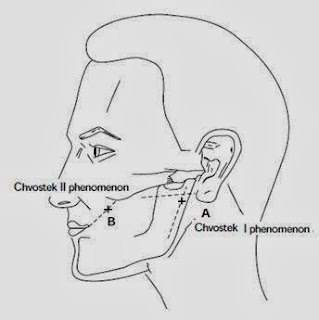
➧ Chvostek's sign: (Figure 1, Figure 2)

(Tap on facial n. anterior to the earlobe or between the zygomatic arch and angle of the mouth → Unilateral spasm of facial muscles)

Chvostek's sign
Figure 1: Chvostek's sign

Chvostek's sign
Figure 2: Chvostek's sign

➧ Trousseau's sign: (Figure 3) 
(Inflate BP cuff 20 mmHg > SBP → Carpopedal spasm)

Trousseau's sign
Figure 3: Trousseau's sign

➧ Hypotension

➧ Bradycardia

➧ Arrhythmias

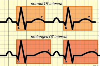
➧ Prolonged QT interval (Figure 4)

Prolonged QT interval
Figure 4: Prolonged QT interval

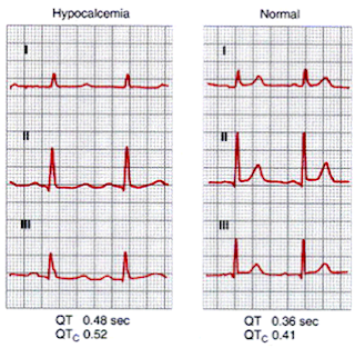
ECG Changes: (Figure 5)


ECG changes in Acute Hypocalcemia
Figure 5: ECG changes in Acute Hypocalcemia

Biochemical Workup:

➧ S total Ca⁺², Albumin and Ionized Ca⁺² 

➧ S PO4⁺² 

➧ S Mg⁺² 

➧ Plasma PTH

- Low in hypoparathyroidism

- High in hungry bones syndrome

➧ 25 (OH)D3 and 1,25 (OH)D3 

➧ S. Amylase and Lipase

Management of Hypocalcemia:

1- First correct low Mg⁺²

2- Control of Tetany:

➧ Calcium gluconate: 10 ml of 10% solution IV over 5-10 min. and repeat as necessary in cases with frank generalized tetany.

➧ Slower continuous infusion of Calcium in less acute cases:

- 10% calcium chloride, 8 ml or 10% calcium gluconate, 22 ml in 100 ml isotonic saline over 10 min.-then continuous infusions of 1-2 mg/kg/h elemental calcium, lasting 6-12 h.; Oral daily maintenance 2-4 g.

- Vitamin D: 1-3 mg/d. oral.

3- Correction of alkalosis:

➧ Isotonic saline.

➧ Ammonium chloride: 2 g/4 h. oral to stop tetany.

Read more ☛ Acute Hypercalcemia

Acute Hypercalcemia

Acute Hypercalcemia

Most Common Causes:

1-Endocrine:

➧ Hyperparathyroidism

➧ MEN

➧ PTH-related peptide (PTHrP) by solid tumors

2-Neoplastic:

➧ Ca with bone metastases

➧ Myeloma

3-Granulomatous:

➧ Sarcoidosis 

➧ Tuberculosis

Clinical picture:

➧ History of polyuria and polydipsia 

➧ Dehydration

➧ Bone pain

➧ Confusion

➧ Anorexia 

➧ Constipation

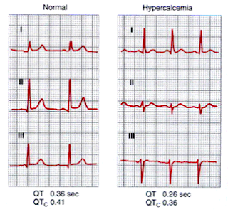
ECG Changes: (Figure 1)


ECG changes in Acute Hypercalcemia
Figure 1: ECG changes in Acute Hypercalcemia

Workup:

S – Ca (High) ➔ PTH (High) ➜ Primary Hyperparathyroidism

➔ PTH (Low) ➜ Malignancy or other cause

S – Ca > 3.0 is 90% of the time of malignant origin

Management of Hypercalcemia:

Volume repletion and diuresis:

➧ NaCl 0.9%: 4 L in first 24 h.

➧ Loop diuretics: Furosemide: 40-80 mg/2 h. IV

-Natriuresis promotes calcium excretion.

Bisphosphonates IV: (Pamidronate /Zoledronate)

-Potent inhibitors of bone resorption.

Corticosteroids:

➧ Prednisone: 30-60 mg/d.

➧ Hydrocortisone: 200mg/d. IV

-Impeding growth of lymphoid neoplastic tissue & enhancing vit. D actions.

Calcitonin:

-4 units/kg/12 h. IM/SC

-Inhibits bone resorption.

Plicamycin-antineoplastic:

-Inhibits resorption.

Dialysis:

-Patients with renal failure.

Urgent Parathyroidectomy


Read more ☛ Acute Hypocalcemia