Educational Blog about Anesthesia, Intensive care and Pain management

Showing posts with label Airway. Show all posts

Airway Blocks

Airway Blocks

1-Superior laryngeal n. block:

Block of the superior laryngeal nerve can provide anesthesia of the larynx from the epiglottis to the level of the vocal cords. This block may be appropriate for any patient requiring TI before anesthetic induction.

Anatomy (Fig. 1):

The superior laryngeal n. is a branch of the vagus n. After it leaves the main vagal trunk, it courses through the neck and passes medially, caudal to the greater cornu of the hyoid bone, at this point, it divides into an internal and external branch. The internal branch is the nerve of interest in the superior laryngeal n. block, and it is blocked where it enters the thyrohyoid membrane inferior to the caudal aspect of the hyoid bone.

Technique (Fig. 1):

The patient is placed supine, with the neck extended. The anesthesiologist should displace the hyoid bone toward the side to be blocked by grasping it between the index finger and the thumb. A 25-gauge, short needle is then inserted to make contact with the greater cornu of the hyoid bone. The needle is walked off the caudal edge of the hyoid and advanced 2 to 3 mm so that the needle tip rests between the thyrohyoid membrane laterally and the laryngeal mucosa medially. 2-3 ml of lidocaine 0.5% is then injected; an additional 1 ml is injected while the needle is withdrawn.

Fig. 1: Superior Laryngeal n. Block


2-Glossopharyngeal n. block:

Glossopharyngeal n. block is useful for anesthesia of the mucosa of the pharynx and soft palate and for eliminating the gag reflex that results when pressure is applied to the posterior third of the tongue, even after adequate topical mucosal anesthesia has been obtained. Glossopharyngeal n. block can be used in most patients who need atraumatic, sedated, spontaneously ventilated, "awake" TI.

Anatomy:

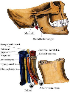
The glossopharyngeal n. exits from the jugular foramina at the base of the skull, in close association with other structures of the carotid sheath, vagus n., and styloid process. It descends in the neck, passes between the internal carotid and the external carotid arteries, and then divides into pharyngeal branches and motor branches to the stylopharyngeus muscle as well as branches innervating the area of the palatine tonsil and the posterior third of the tongue. These distal branches of the glossopharyngeal n. are located submucosally immediately posterior to the palatine tonsil, deep to the posterior tonsillar pillar.

a) Intraoral approach (Fig. 2):

After topical anesthesia of the tongue, the patient's mouth is opened widely, and the posterior tonsillar pillar (palatopharyngeal fold) is identified by using a No. 3 Macintosh laryngoscope blade. Then an angled 22-gauge, 9-cm needle, (this can be done by using a 22-gauge disposable spinal needle. In an aseptic manner, the stylet is removed from the disposable spinal needle and discarded. Subsequently, the distal 1 cm of the needle is bent to allow more control during submucosal insertion) and is inserted at the caudad portion of the posterior tonsillar pillar. The needle tip is inserted submucosally, and then, after careful aspiration for blood, 5 ml of lidocaine 0.5% is injected. The block is then repeated on the contralateral side.

Fig. 2: Glossopharyngeal n. Block (Intraoral approach)


b) Peristyloid approach (Fig. 3):

The patient lies in a supine position, with the head in a neutral position. Marks are placed on the mastoid process and the angle of the mandible. A line is drawn between these two marks, and at the midpoint of that line, the needle is inserted to contact the styloid process. To facilitate styloid identification, a finger palpates the styloid process with deep pressure, and, although this can be uncomfortable for the patient, the short 22-gauge needle is then inserted until it impinges on the styloid process. This needle is then withdrawn and redirected off the styloid process posteriorly. As soon as bony contact is lost and aspiration for blood is negative, 5-7 ml of lidocaine 0.5% is injected. The block can then be repeated on the contralateral side.

Fig. 3: Glossopharyngeal n. Block (Peristyloid approach)


3-Translaryngeal Block:

Anatomy (Fig. 4):

Translaryngeal block is most useful in providing topical anesthesia to the laryngotracheal mucosa innervated by branches of the vagus nerve. Both surfaces of the epiglottis and laryngeal structures to the level of the vocal cords receive innervation through the internal branch of the superior laryngeal nerve, a branch of the vagus. The distal airway mucosa also receives innervation through the vagus nerve but via the recurrent laryngeal nerve. Translaryngeal injection of local anesthetic helps provide topical anesthesia for both these vagal branches, since injection below the cords through the cricothyroid membrane results in solution being spread onto the tracheal structures and coughed onto the more superior laryngeal structures.

Technique (Fig. 4):

The patient should be in a supine position, with the pillow removed and the neck slightly extended. The anesthetist should be in a position to place the index and third fingers in the space between the thyroid and the cricoid cartilage (cricothyroid membrane). The cricothyroid membrane should be localized, the midline identified, and the needle, 22-gauge or smaller, inserted into the midline until air can be freely aspirated. When air can be freely aspirated, 3-4 mL of 4% lidocaine is rapidly injected. The needle should be removed immediately since it is almost inevitable that the patient will cough at this point. Conversely, a needle-over-the-catheter assembly (intravenous catheter) can be used for the block. Once air has been aspirated, the inner needle is removed, and the injection is performed through the catheter.

Fig. 4: Translaryngeal Block


Potential Problems:

This block can result in coughing, which should be considered in patients in whom coughing is clearly undesirable. The midline should be used for needle insertion since the area is nearly devoid of major vascular structures. Conversely, the needle does not need to be misplaced far off the midline to encounter significant arterial and venous vessels.

Laryngeal Mask Airway

Laryngeal Mask Airway (LMA)

1-The LMA-Classic™:


-It is a reusable LMA™ airway for general anesthesia.
-The LMA-Classic™ is available in eight sizes: (1, 1½, 2, 2½, 3, 4, 5, and 6).

Advantages:

-A safe and effective alternative to the endotracheal tube and the facemask.

-Over 100 million uses worldwide.

-Leaves the anesthetist's hands free to attend, to monitoring and record keeping.

-Latex-free and exceptionally well tolerated.

-A reusable device that can be cleaned and steam sterilized up to 40 times before being discarded.

2-LMA-Unique™ (Single-use):


-The LMA-Unique™ is a convenient, single-use LMA™ airway suitable for general anesthesia procedures.

-The LMA-Unique™ is available in sizes: (1, 1½, 2, 2½, 3, 4, and 5).

-The LMA-Unique™ is a disposable, single-use device, made to the same design specifications as the LMA-Classic™.

Advantages:

-Packaged sterile - ready for use.

-Suitable for use on emergency vehicles.

-Suitable where access to sterilization facilities is limited.

-Made of medical-grade PVC.

3-LMA-Flexible™:


-The LMA-Flexible™ is a reinforced LMA™ airway with a flexible airway tube.

-The LMA-Flexible™ is available in sizes: (2, 2½, 3, 4, 5, and 6).

-The LMA-Flexible™ is a re-usable device that can be cleaned and steam sterilized up to 40 times before being discarded.

Advantages:

-Designed for ENT, dental, and head surgery.

-Allows extreme flexion.

-Guaranteed kink and crush-proof.

-Latex-free.

4-LMA Flexible (Single-use):


-The LMA Flexible™ Single Use is ideal for use in ENT, ophthalmic, dental, and other head and neck cases and extends the LMA™ Airway benefits of hemodynamic stability and smoother emergence to more procedures.

5-LMA-ProSeal™:


-The LMA-ProSeal™ is an advanced LMA™ airway suitable for general anesthesia.

-It has a unique double cuff arrangement that provides an exceptionally effective, 'hands-free' airway seal, at low intracuff pressures.

-The LMA-Proseal™ is available in sizes: (1½, 2, 2½, 3, 4, and 5).

-The LMA-Proseal™ is a re-usable device that can be cleaned and steam sterilized up to 40 times before being discarded.

Advantages:

-A new double tube design separates the respiratory and alimentary tracts, providing a safe escape channel for regurgitated fluids in the event of unexpected regurgitation.

-The mask is designed to be a minimally stimulating airway device, whose cuff tip presses against the upper oesophageal sphincter when it is correctly positioned. The sides of the mask face the pyriform fossae and the upper border rests against the base of the tongue.

-Latex-free.

6-LMA Supreme™ (Single-use):


-The first and only single-use laryngeal mask with a built-in drain tube.
-The integrated drain tube is designed to channel fluid and gas safely away from the airway. Several simple and quick tests help verify accurate positioning.
-An improved curve for easy insertion. Subtle refinements in the mask make correct placement easier.

7-LMA Fastrach™ & LMA Fastrach™ ETT:


-The design of the LMA-Fastrach™ facilitates rapid insertion from any position, even if space is limited, and moving the patient is a possible hazard.

-The device is self-positioning with the rigid tube designed to fit the curvature of the palatopharyngeal arch, enabling a firm seal to be achieved.

-The LMA-Fastrach™ is available in sizes: (3, 4, and 5).

-The LMA-Fastrach™ is a reusable device that can be cleaned and steam sterilized up to 40 times before being discarded.

Advantages:

The LMA-Fastrach™ has additional features to those of the LMA-Classic™:

-Designed specifically for the anatomically difficult airway.

-Ideal in emergency situations.

-Can be used as an intubating tool, with no interruption of patient oxygenation.

-Allows insertion in the neutral position, in limited space.

-No need to move the patient.

-No need to insert fingers into the patient's mouth.

8-LMA Fastrach™ & LMA Fastrach™ ETT (Single-use):

9-LMA CTrach™:


-The only difficult airway device that allows ventilation, visualization, and intubation.

-The LMA CTrach™ is designed to increase intubation success rates in difficult airways. The LMA CTrach™ mask enables ventilation during intubation attempts while built-in fiber optics provide a direct view of the larynx and real-time visualization of the ET tube passing through the vocal cords.

-The LMA CTrach™ can be inserted exactly the same as the LMA Fastrach™, however, unlike the LMA Fastrach™, once the airway is secured and the patient is being ventilated, the viewer is switched on, placed in the magnetic connector, and a clear image of the larynx is displayed in real-time. The ET tube can be viewed as it enters the trachea. Once the patient is intubated, the viewer is removed and the mask is removed leaving the ET tube in place.

10-i-gel™:


-The i-gel supraglottic airway device accurately and naturally positions itself over the laryngeal framework to provide a reliable peri-laryngeal seal without the need for an inflatable cuff.

-i-gel is made from a medical-grade thermoplastic elastomer, i-gel has been designed to create a non-inflatable, anatomical seal of the pharyngeal, laryngeal, and peri-laryngeal structures whilst avoiding compression trauma.

-i-gel is currently available in sizes: (1, 1½, 2, 2½, 3, 4, and 5), and is supplied in an innovative, color-coded polypropylene ‘cage pack’.

Advantages:

-No inflatable cuff offers easy, rapid insertion.

-An integral bite block reduces the possibility of airway occlusion.

-A buccal cavity stabilizer aids rapid insertion and eliminates potential rotation.

-Made from a unique, soft, gel-like material to allow easy insertion and reduced trauma.

-Gastric channel designed to improve and enhance patient safety.

-Reduces the possibility of epiglottis downfolding and obstructing the airway.

-Unique packaging protects the i-gel in transit and ensures maintaining of its anatomical shape.

Traumatic Complications of TI

Traumatic Complications of TI


Larynx
➧ Despite the frequent use of tracheal intubation (TI) for both short-term events like surgery or for long-term ventilatory support, traumatic complications from TI still occur.

➧ Both the upper and lower airways are at risk of injury. The upper airway includes the naso- and oro-pharynx and extends to the level of the vocal cords. The lower airway lies distal to the vocal cords. 


1-Lips injury:

➧ Frequently the right lower lip is caught between the laryngoscope blade and the lower teeth. 

➧ It can also occur when the endotracheal tube (ETT) is placed or when a hard plastic oral airway is placed. 

➧ There is frequently soft tissue injury to the lip, but serious consequences are rare.

2-Dental injury:

➧ The upper incisors are usually involved. Identified risk factors include preexisting poor dentition and one or more indicators of difficult laryngoscopy and intubation. 

➧ A protective plastic shield placed over the upper teeth may limit the potential for damage to teeth.

3-Tongue injury:

➧ Prolonged compression by an ETT, an oral airway, or both may impair circulation, leading to ischemia and poor venous drainage, which then leads to macroglossia. 

➧ Obstruction of the submandibular duct by an ETT may lead to massive tongue swelling. 

➧ Compression injury to the lingual n. during difficult intubation has been reported with a one-month loss of tongue sensation.

4-Uvular injury:

➧ Injury to the uvula is a rare complication of TI and is usually associated with mechanical interference with the blood supply to the uvula during intubation. 

➧ Common causes of such interference include compression caused by excessive length of the ETT leading to direct pressure, blind pharyngeal suction with a hard suction catheter, and entrapment of the uvula between the ETT and oral airway. 

➧ This condition causes sore throat, difficulty in breathing, painful swallowing, and foreign body sensation in the throat. 

➧ Treatment of patients with steroids and antibiotics is successful, with complete healing in 2 weeks.

5-Nasal injury:

➧ Nasal intubations in particular are associated with mucosal tears or lacerations that may result in significant epistaxis. 

➧ The use of nasal decongestants/vasoconstrictors, lubrication of the ETT, and warming of the tube before nasal intubation may reduce the risk of epistaxis. 

➧ Once the nasotracheal tube is in place, it is important to position the tube in such a way as to prevent distortion of the nares, which can lead to local ischemia, followed by necrosis. 

➧ Blind nasal intubation also increases the risk of oropharyngeal and laryngeal injury, as does oral intubation with a protruding stylet. 

➧ Pharyngeal injury can progress into retropharyngeal infections or mediastinitis, and positive pressure ventilation may produce subcutaneous emphysema, pneumomediastinum, and pneumothorax.

6-Temporal-Mandibular Joint injury:

➧ This joint can be dislocated during TI.

7-Cervical Spine injury:

➧ Risk factors for spinal injury include head and neck trauma, cervical osteoporosis, atlantoaxial instability as seen in patients with rheumatoid arthritis and Down's syndrome, and lytic bone lesions. 

➧ Immobilization of the head and neck will prevent most spine injuries regardless of the route or method of intubation.

8-Pharyngeal and Esophageal injury:

➧ Venous drainage from the pharynx may be impaired by the mechanical presence of the tracheal tube and result in pharyngolaryngeal edema. 

➧ Both pharyngeal and esophageal perforations can occur after a difficult TI and in patients older than 60 y.

9-Laryngeal injury:

a) Vocal Cord Paralysis:

➧ Vocal cord paralysis is attributed to nerve injury or mechanical injury. 

➧ It may be unilateral or bilateral, bilateral injury is riskier and frequently requires emergency reintubation or tracheotomy. 

➧ The mechanism may be related to inflation of the ETT cuff at the level of the subglottic larynx rather than at the correct location in the trachea where an anterior branch of the recurrent laryngeal n. enters between the cricoid and the thyroid cartilage, providing innervation to the intrinsic muscles of the larynx. 

➧ An inflated cuff at this location can compress the nerve between the cuff and the overlying thyroid cartilage, causing injury. 

➧ This can be exacerbated when N₂O is used as part of the general anesthesia, as it diffuses rapidly into the cuff, increasing cuff pressure (Pcuff) and risk for injury. 

➧ Vocal cord immobility may also be caused by pseudolaryngeal paralysis which can be associated with cricoarytenoid dislocation, arytenoid fusion, and posterior glottic stenosis. 

➧ Interarytenoid fibrous adhesion can occur after intubation and is frequently confused with bilateral vocal cord paralysis. 

➧ Measures to decrease the risk for recurrent laryngeal n. injury include: 

-Use of a low-pressure, high-volume ETT cuff 

-Avoidance of larger than necessary ETTs 

-Avoidance of overinflation of the ETT cuff 

-Prevention of excessive tube migration during anesthesia 

➧ Vocal cord paralysis is usually associated with spontaneous recovery.

b) Arytenoid Cartilage Dislocation:

➧ The arytenoid cartilage can be dislocated from the cricoarytenoid joint due to the pressure of the tip of the tracheal tube as it is placed through the vocal cords. 

➧ This rare complication can occur with routine elective intubation and most often during traumatic intubation, intubations in which the patient is not relaxed and is struggling or coughing during tube placement and with the use of certain airway devices such as the McCoy laryngoscope or the lighted stylet (lightwand). 

➧ Acutely dislocated arytenoid cartilage can be reduced using rigid bronchoscopy. 

➧ If the dislocation is not reduced, chronic hoarseness and vocal cord dysfunction often result.

c) Hematoma and Granuloma formation:

➧ Hematoma formation on the vocal cords has been noted after routine TI and usually resolves without sequelae. 

➧ Occasionally scarring of the injured area occurs, resulting in a chronically hoarse voice. 

➧ Granulomas can also form after hematomas on the vocal cords resulting from TI. 

➧ Granuloma formation after intubation has been described as occurring with an incidence of 1:800 to 1:20000 in adults. 

➧ The most common site is the vocal process of the arytenoids because this structure comes into close contact with the ETT. 

➧ The degree of injury increases with increasing tube size and duration of intubation.

10-Tracheal injury:

➧ Tracheal laceration as a result of intubation was first reported in 1977. 

➧ Factors related to this type of injury include: 

-Overinflation of the ETT cuff 

-Multiple intubation attempts 

-Use of stylets 

-Malpositioning of the tube tip 

-Tube repositioning without cuff deflation 

-Inadequate tube size 

-Vigorous coughing 

-N₂O in the cuff 

-Traumatic intubation 

-Emergency intubation 

-Anesthesia protocol 

-An oversized tube 

-Long duration of intubation. 

➧ The risk is also greater in patients with: 

-Tracheal distortion caused by neoplasm or large lymph nodes 

-Weakness in the membranous trachea (seen in women or the elderly) 

-Chronic obstructive lung disease 

-Corticosteriod therapy. 

➧ The most common site for tracheal rupture is the junction of the cartilage and posterior membrane on the right side and the length of the tear often corresponds to the length of the cuff on the ETT. 

➧ Translaryngeal ETTs can cause circumferential stenosis or malacia at the cuff site. Tracheal stenosis can develop after even a brief (<24 h.) TI. 

➧ The incidence of injury at the cuff site was likely higher in the era of high-pressure, low-volume cuffs and has certainly declined with the use of low-pressure, high-volume cuffs since the 1970s. 

➧ The newer cuffs, however, can easily be overinflated, exceeding the tracheal mucosal capillary perfusion pressure of 20-30 mmHg, and resulting in local tissue ischemia, which is directly proportional to the tracheal tube Pcuff. (Methods to stabilize ETT intracuff pressure)

➧ At a Pcuff of 30 cmH₂O, the tracheal mucosal blood flow becomes partially obstructed, and at a pressure of 45 cmH₂O, the obstruction becomes total, leading to tracheal mucosal damage and subsequent complications. 

➧ These lesions heal with either fibrous stenosis or loss of cartilaginous support and ensuing malacia.

Diagnosis:

➧ The clinical manifestations of laryngeal and tracheal injury can range from mild hoarseness to severe stridor. 

➧ Patients also may present with other non-specific symptoms such as exertional dyspnea and positional wheezing. 

➧ The airway is typically narrowed to less than 8 mm before exertional dyspnea is present. 

➧ Once the lumen is less than 5 mm, symptoms of dyspnea and stridor may present at rest. 

➧ Due to the dramatic loss of airway diameter before the development of symptoms, up to 54% of patients with tracheal stenosis can present in respiratory distress.

Treatment:

➧ Treatment of airway injury from prolonged intubation varies with the cause of injury but may include: 

-Bronchoscopy and dilation 

-Laser treatments of granulomas and webs 

-Resection of stenotic tracheal rings 

-Splitting the cicoid gland (cartilage).


Complications while ETT in place

Complications while ETT is in place

A) Obstruction of the endotracheal tube (ETT):

Causes:

1-Accumulation of secretions in the tube 

2-Kinking of the tube.

B) Misplacement or migration of the ETT:

➧ A common complication and right mainstem intubation has been associated with increased mortality in critically ill patients. 

➧ Traditional methods of assuring proper tube position include: 

-Observing bilateral chest expansion.

-Auscultation of bilateral breath sounds and the lack of air in the epigastrium. 

➧ Unfortunately, none of these methods is reliable, leading to the use of capnography as the current gold standard to detect placement of the tube in the airway and rule out esophageal placement. 

➧ Capnography, however, does not detect proximal or distal placement of the ETT within the airway.

C) Respiratory Complications of TI:

1-Dead Space

➧ Tracheal intubation usually results in a small reduction in dead space ventilation (25-75 ml); this reduction is too small to be beneficial in most patients.

2-Airflow Resistance

➧ Resistance to airflow through ETT is significant, particularly at small tube diameters. 

➧ Most airflow through ETTs is turbulent. The resistance to flow is therefore proportional to the fifth power of the radius. This can result in marked differences in the pressure necessary to cause airflow depending on the internal diameter of the ETT and the flow rate required.

3-Airway Resistance

➧ Intubation also causes an increase in the lower airway resistance in most people as a result of parasympathetic activation of airway smooth muscle. Generally, the increase in lower airway resistance is not clinically significant.

4-Bronchospasm

➧ TI can trigger severe bronchospasm. Interestingly, only one-half of the patients in whom bronchospasm occurred had a previous history of bronchial asthma or pulmonary disease. 

➧ Preoperative inhaled β-agonists such as albuterol as well as anticholinergic agents may prevent bronchospasm in patients with reactive airway disease. 

➧ Inhaled/IV β-agonists, anticholinergic agents such as glycopyrrolate, and an increase in the level of inhaled anesthetic agents; all have been used intraoperatively to treat bronchospasm. 

➧ In some instances, the tracheal tube must be removed before the bronchospasm will stop.

5-Cough

➧ ETT results in reduced cough efficiency and, if humidification is not provided, drying of the upper airway.

6-PEEP

➧ The presence of ETT through the glottis reduces the small positive pressure or "auto-PEEP" caused by the resistance of air flowing through the glottis. This loss of PEEP may reduce the functional residual capacity in some patients in respiratory failure dependent on "auto-PEEP" for oxygenation.

7-Respiratory Mucosa

➧ The presence of an artificial airway leads to functional and morphologic changes in the respiratory mucosa due to the loss of the humidification, warming, and filtering effects of the upper airway (especially the nasopharynx). In the long term, this may result in squamous metaplasia and granulation tissue formation.


Hemodynamic effects of Laryngoscopy and TI

Hemodynamic effects of Laryngoscopy and TI


Autonomic innervation and response of the airway:

➧ The area of the trachea and pharynx is richly innervated and involves both the parasympathetic and sympathetic nervous systems.

➧ Following the mechanical stimulation of the upper respiratory tract (URT) (i.e. nose, epipharynx, laryngopharynx), the afferents are carried by the glossopharyngeal nerve and from the tracheobronchial tree via the vagus nerve which enhances the activities of the cervical sympathetic afferent fibers resulting in a transient rise in heart rate (HR) and blood pressure (BP). 

➧ The lower respiratory tract (LRT) is protected by reflex arcs from both the upper and lower airways. The afferent pathways are comprised of the glossopharyngeal nerves in the oropharynx, superior to the anterior surface of the glottis, and the superior and recurrent laryngeal nerves for the posterior and inferior glottis, whereas the efferent pathway is controlled by the vagus. 

➧ Afferent stimuli can therefore trigger cardiac, airway, cerebral, neuromuscular, and adrenal responses. 

➧ The hemodynamic responses to orotracheal intubation have two components. The first is the response to laryngoscopy and the second is the response to tracheal intubation (TI). 

➧ Tachycardia and hypertension have been reported since 1950 during intubation under light anesthesia as TI causes a reflex increase in sympathetic activity. 

➧ The hemodynamic responses are due to reflex sympathoadrenal discharge provoked by epilaryngeal and laryngotracheal stimulation after laryngoscopy and TI, this results in hypertension, tachycardia, arrhythmia, and a change in plasma catecholamine concentrations. 

➧ Translaryngeal intubation of the trachea stimulates laryngeal and tracheal receptors, resulting in a marked increase in the elaboration of sympathomimetic amines. This sympathetic stimulation results in tachycardia and a rise in BP. 

➧ In normotensive patients, this rise is approximately 20-25 mmHg; it is much greater in hypertensive patients. Nasopharyngeal intubation causes a significant pressor response. 

➧ Stimulation of the larynx and trachea by the passage of the tracheal tube, but not direct laryngoscopy, causes a significant increase in this response. Direct stimulation of the trachea appears to be a major cause of the hemodynamic changes associated with TI. 

➧ The extent of the reaction is affected by many factors: the technique of laryngoscopy and intubation, and the use of various airway instruments. Laryngoscopy itself is one of the most invasive stimuli during orotracheal intubation. 

➧ Many anesthesiologists agree that applying a small force to the patient’s larynx when using a laryngoscope might prevent excessive hyperdynamic responses to orotracheal intubation. 

➧ In infants, laryngoscopy and TI often result in bradycardia from vagal stimulation. Administration of anticholinergic agents such as atropine can block this response. In older children or adults, this vagal response is rarely observed. Although bradycardia can develop in up to 10% of patients undergoing TI, the typical result is tachycardia and hypertension, leading to an increase in myocardial oxygen consumption. It has been shown that up to 15% of patients undergoing TI under general anesthesia will have ventricular arrhythmias, with the majority of events occurring at the time of tube insertion, as opposed to at the time of laryngoscopy. 

➧ The rise in HR and BP occurs about 14 sec. after the start of direct laryngoscopy and becomes maximal after 30-45 sec. Prolonged intubation time in difficult airways, in addition, induces hypercarbia and decreases anesthetic gas concentration, resulting in tachycardia and hypertension. These responses are usually transient and innocuous.

Cardiac Patients:

➧ In patients with co-existing hypertension or ischemic heart disease, these may be exaggerated or may jeopardize the balance between myocardial oxygen requirements and delivery. In these patients, it is important to minimize the duration of direct laryngoscopy, if possible, to less than 15 sec. 

➧ During and immediately following TI associated with tachycardia and hypertension, there is a decrease in the left ventricular ejection fraction (stroke volume/end-diastolic volume). This is particularly marked in patients with coronary artery disease. 

➧ Increases in HR may be associated with ST-segment changes that indicate myocardial ischemia. 

➧ The cardiovascular response to TI can be problematic if the patient suffers from cardiac disease, cerebrovascular or abdominal-vascular disease in which hypertension may lead to hemorrhage.

Hypertensive Patients:

➧ Hypertensive patients are prone to greater and exaggerated circulatory responses after laryngoscopy and TI, because of long-term persistent vascular hyperreactivity, than normotensive patients. An increase in BP associated with TI is dangerous and may cause complications, including pulmonary edema, heart failure, and cerebrovascular hemorrhage. Therefore, prevention of these pressor responses is of particular importance in hypertensive patients.

Elderly Patients:

➧ Transient tachycardia and hypertension associated with laryngoscopy and TI are probably of little consequence in young healthy patients, but either or both may be hazardous to elderly patients, especially to those with hypertension or myocardial insufficiency. Elderly patients have a high incidence of clinical and occult coronary artery disease, and age is a major risk factor for perioperative cardiac morbidity. This risk may be minimized by the maintenance of a balance between myocardial oxygen supply and demand. Thus, the maintenance of hemodynamic stability during TI is of particular clinical importance in elderly patients with hypertension.

Intracranial Pressure (ICP):

➧ Sympathetic stimulation from TI also increases ICP; this can be harmful in patients with intracranial mass lesions or increased ICP from other pathology. The patient with elevated ICP who has a minimum reserve in intracranial compliance is actually at risk for brain-stem herniation and sudden death during laryngoscopy and TI. Instrumentation of the airway may result in a sudden increase in cerebral blood flow due to increases in cerebral metabolic activity and systemic cardiovascular effects. The normal autoregulation mechanism may not be effective because of disease or because its upper-pressure limit (normally, mean arterial pressure 150 mmHg) may be exceeded. Coughing or bucking will decrease venous return from the head and may increase ICP as well.

Intraocular Pressure (IOP):

➧ The mechanism of IOP rise is secondary to increased sympathetic activity. Adrenergic stimulation causes vaso- and veno-constriction, and an increase in central venous pressure, which has a close relationship with IOP. 

➧ In addition, adrenergic stimulation can also produce an acute increase in IOP, by increasing the resistance to the outflow of aqueous humor in trabecular meshwork between the anterior chamber and Schlemm’s canal. 

➧ The acute increase in IOP may be dangerous for patients with impending perforation of the eye, perforating eye injuries, and glaucoma. 

➧ Control of IOP during ophthalmic surgery or diagnostic tonometry is clinically important, because uncontrolled IOP increases induced by airway manipulation may worsen ocular morbidity or produce misleading results.


Read more ☛ about Traumatic Complications of TI

Methods to stabilize ETT intracuff pressure

Methods to stabilize ETT intracuff pressure

-The use of N₂O, which is well-known to diffuse into ETT cuffs, and the lack of frequent control of intracuff pressure (iPcuff) are the most important factors that contribute to the high incidence of excessive iPcuff during the perioperative period. Other factors, such as the diffusion of O₂ into the cuff and the warming of gases inside the cuff, play a small role in the increase in the iPcuff. Various factors affect the rate of diffusion of N₂O, including the difference in partial pressure of N₂O inside and outside the cuff, the area available for diffusion, and the cuff material. Prevention of overpressure of the ETT cuff can be achieved by several means:

1-Manual palpation of the pilot balloon:

-Free inflation of the cuff, controlled only by the anesthesiologist’s manual palpation of the pilot balloon, is not reliable and results in extremely variable iPcuff, ranging from 0-120 cmH₂O.

2-The pinch test:

-The pinch test represents an extension of the practice of palpating the pilot balloon of an ETT to verify the proper inflation of the cuff. The pilot balloon of the ETT is compressed manually between the anesthesiologist’s thumb and index finger until it is flat and the time to its reinflation is measured using a stopwatch. The regression analysis allowed the following equation to be developed for the restoration time (T) in sec. as a function of the (Pcuff) in cmH₂O: [T = 2.72 − 0.041 * Pcuff].

3-Simple on-line relief valve:

-It is a simple and inexpensive method to gauge the Pcuff by using a regular 20-ml syringe attached in line with the connector of the ETT cuff. The syringe is connected to the tube cuff and inflated with 15 ml of air and is left constantly connected to the cuff. This results in an adequate venting of the excess iPcuff and also there is no leakage around the cuff.

4-Automatic regulation of the cuff pressure:

-The procedure requires only a simple aquarium air pump and conventional tubing. The procedure devised to maintain ETT Pcuff is readily implemented, cheap, easy to operate and can be used regardless of the specific ventilator or tube used.

5-The Pcuff monitoring devices:

a) ETT Intracuff Pressure Manometer: (Figure 1)


ETT Intracuff Pressure Manometer
Figure 1: ETT Intracuff Pressure Manometer

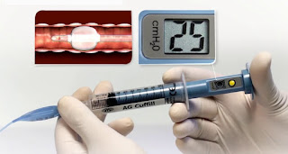
b) AG Cuffill Cuff Inflator with Integrated Manometer: (Figure 2)


AG Cuffill Cuff Inflator with Integrated Manometer
Figure 2: AG Cuffill Cuff Inflator with Integrated Manometer

c) Tru-Cuff ETT Cuff Pressure Syringe: (Figure 3)


Tru-Cuff ETT Cuff Pressure Syringe
Figure 3: Tru-Cuff ETT Cuff Pressure Syringe

d) Vortan Cuff Inflator: (Figure 4)


Vortan Cuff Inflator
Figure 4: Vortan Cuff Inflator

e) AccuCuff Cuff Pressure Indicator: (Figure 5)


AccuCuff Cuff Pressure Indicator
Figure 5: AccuCuff Cuff Pressure Indicator

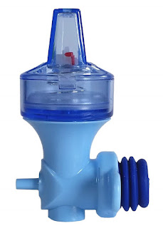
f) PressureEasy Cuff Pressure Controller: (Figure 6)


PressureEasy Cuff Pressure Controller
Figure 6: PressureEasy Cuff Pressure Controller

6-Profile Soft-Seal Cuff (PSSC) ETT:

-The Profile Soft-Seal Cuff (PSSC; Sims Portex, Kent, UK), made of a material impervious to N₂O, velvet soft polyvinyl chloride, a new material with N₂O gas barrier properties produces a thin and highly compliant cuff without increasing N₂O diffusion, thereby reducing the increase of iPcuff and postoperative sore throat.

7-Portex Soft-Seal tube cuff:

-In the Portex Soft-Seal tube cuff (Portex Ltd., Hythe, UK), the plasticizer added to soften the polyvinyl chloride makes the cuff much less permeable to N₂O despite having a thickness of 0.06 mm, which is very similar to the Mallinckrodt Lo-Contour (Athlone, Ireland). The new design prevented increases in the iPcuff and remained stable.

8-Repeated deflation of Air-filled ETT cuff:

-In clinical practice, some anesthesiologists perform a simple method of repeated cuff deflations to inhibit excessive pressure, and eventually, the Pcuff stabilizes. Immediately after TI, the ETT cuff is aspirated as much as possible and then inflated with the smallest volume of air that would produce 12-14 mmHg of Pcuff and seal the airway when the intra-airway plateau pressure is 18 cmH₂O.

-During 67% N₂O anesthesia, gases in the cuff are aspirated every 30 min. in the Trachelon (Terumo, Tokyo, Japan) or 60 min. in the PSSC ETT (Sims Portex, Kent, UK) for 4 h. to decrease the Pcuff to the initial pressure. Pcuff should stabilize when the N₂O concentration is equivalent on both sides of the cuff wall. Therefore, equilibration of the N₂O concentration after repeated deflation for 4 h. is the underlying mechanism of this simple technique. The increased compliance of the PSSC ETT cuff, rather than the N₂O gas barrier, contributes to the requirement for longer intervals between cuff aspirations to avoid excessive pressure compared with what is needed for standard ETTs.

9-N₂O-O₂ gas mixture-filled ETT cuff:

-Immediately after intubation, the cuff is aspirated as much as possible and then inflated with the smallest volume of 40% N₂O and 60% O₂ that would not leak when the intra-airway plateau pressure is 18 cmH₂O. The N₂O gas mixture to fill cuffs is aspirated from the common gas outlet of an anesthetic machine.

-Inflating cuffs with 40% N₂O maintains stable Pcuff without excessive iPcuff or air leaks during anesthesia with 67% N₂O; however, deflationary phenomena of the cuff might occur because iPcuff decreases so quickly after substituting oxygen for N₂O, therefore, it is suggested that the Pcuff be checked frequently to avoid air leaks and aspiration of gastric contents during prolonged emergence from anesthesia or during transportation of patients with TI. 

-The ETT with the N₂O gas-barrier type of cuff might be beneficial because of the longer time required to decrease Pcuff after cessation of N₂O administration and that the highly compliant cuff might be the mechanism of slow changes of iPcuff in the PSSC.

10-Saline-filled ETT cuff:

-When saline is used to fill the cuff, the lack of pressure increase secondary to N₂O diffusion depending on the physical principle that liquids do not expand in volume when highly soluble gases dissolve in them.

11-Lidocaine-filled ETT cuff:

-Lidocaine placed inside the cuff of an ETT can slowly diffuse through its hydrophobic structure. The ETT cuff is prefilled with 7 to 8 ml of L-HCl 2% (140-160 mg) for 90 min. before intubation to enhance the diffusion of lidocaine across the cuff then the cuff is evacuated before intubation. Following intubation, the ETT cuff is inflated with enough lidocaine to prevent retrograde leak at a tidal volume of 10 ml/kg.

12-Alkalinized lidocaine-filled ETT cuff:

-Alkalization of lidocaine can promote the in vitro diffusion across the ETT cuff many tens of times. The cuff of the tracheal tube is initially inflated slowly with saline until no leak is heard under controlled ventilation, after the initial injection of 2 ml of L-HCl 2% (40 mg) into the ETT cuff, a supplementary volume of 2 ml of 8.4% NaHCO₃ is added. It has been shown that small amounts of L-HCl diffused slowly across the ETT cuff; the addition of NaHCO₃ increases the diffusion and dramatically increases the amount of lidocaine released. Plasma lidocaine concentrations increase when lidocaine is alkalinized (Cmax is 62.5 ± 34.0 ng/ml) versus (3.2 ± 1.0 ng/ml) without NaHCO₃. Alkalinization of L-HCl with NaHCO₃ allowed the diffusion of 65% (versus 1% without NaHCO₃) of the neutral base form of lidocaine for 6 hours.

-Use of a small dose of alkalinized lidocaine markedly improves ETT tolerance during a more prolonged time and offers the advantages of minimal stress response to smooth extubation and cough-free emergence.