Educational Blog about Anesthesia, Intensive care and Pain management

Showing posts with label GIT diseases. Show all posts

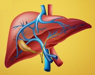
Anesthetic Considerations for Patients with Liver disease

Anesthetic Considerations for Patients with Liver Disease


Preoperative:

1. Assess the Degree of hepatic impairment, Severity, and Hepatic reserve by the Child-Turcotte-Pugh scoring system.

2. AVOID: Premedication, IM injections, Contact with blood or body fluids, unnecessary esophageal instrumentation.

Regional Anesthesia:

-Regional anesthesia might be used when possible in patients with advanced liver disease.

-Coagulopathy (PT & INR) should be considered a contraindication to some types of regional anesthesia.

-AVOID Epidural a. (Large amounts of amide LAs).

IV Anesthetics:

-Propofol, Ketamine (in hypotensive patients).

Opioids:

-Opioids can also be used successfully in patients with the hepatic disease despite certain pharmacological consequences (decreased clearance and prolonged half-life).

-Fentanyl is considered the opioid of choice because it does not decrease hepatic oxygen and blood supply nor does it prevent increases in hepatic oxygen requirements when used in relatively moderate doses.

-AVOID Morphine (Active metabolite, Prolonged action).

Changed Pharmacokinetics:

-The half-life of lidocaine in patients with liver disease may be increased by more than 300%, for benzodiazepines by more than 100%, etc.

-For drugs binding to albumin, the volume of distribution is decreased and therefore the drug dosage should be decreased (e.g. sodium pentothal).

Muscle Relaxants:

-Suxamethonium → Prolonged action, Atracurium, Cisatracurium (of choice).

-AVOID Pancuronium, Vecuronium (Hepatic metabolism).

-The volume of distribution of many drugs can be substantially increased (for different reasons, including an increase in gamma globulin and edema), dictating a necessity to increase the first effective dose of the drug.

-However, owing to a decrease in hepatic blood flow and hepatic metabolic and excretory functions, as well as impaired renal function, the clearance of such a drug is decreased, therefore the effect can be prolonged (e.g. pancuronium).

-Atracurium has a theoretical advantage because its metabolism is not dependent on liver function. Therefore, the clearance and elimination half-life of atracurium in patients with impaired hepatic and/or renal function is not different from those with normal hepato-renal function. However, the volumes of distribution are larger, and, accordingly, the distribution half-lives are shorter in patients with severe hepato-renal dysfunction compared with normal individuals.

-Titration of any relaxant according to the transcutaneous nerve stimulation monitoring is beneficial because the degree of hepatic dysfunction affects the degree of pharmacokinetic disorders.

Inhalational Anesthetics:

-Halothane should be avoided because it leads to the most prominent decrease in hepatic blood flow and oxygen supply and postoperative hepatic dysfunction. In addition, immunologically mediated severe postoperative halothane hepatitis may follow halothane anesthesia.

-Isoflurane is a better choice if an inhalational technique is selected.

-More recently introduced volatile anesthetics, sevoflurane, and desflurane, each of them, can be used safely in patients with liver disease, as they preserve hepatic blood flow.

-Nitrous oxide has been used in patients with advanced hepatic disease for many years, and so far has not been incriminated in increased anesthesia-related hepatic postoperative complications. However, a well-known sympathomimetic effect of nitrous oxide and some possibilities of jeopardizing oxygenation render the routine use of nitrous oxide in patients with advanced liver disease undesirable. It is important to remember that long surgical operations under anesthesia with nitrous oxide might result in the accumulation of nitrous oxide in the intestinal lumen with subsequent intestinal distension.

Others:

-Renal function must be maintained by administering proper fluid load (volume and content); (avoid Na+ overload, use glucose-containing solutions for hypoglycemia, albumin 5% is the preferred colloid), and diuretics if needed.

-The parameters of controlled ventilation should be carefully selected to avoid an unnecessary increase in intrathoracic pressure which may impede venous return thereby decreasing cardiac output.

-Monitoring the coagulation state during surgery can be important. The treatment should be based on the results of hematologic monitoring and may include administration of platelets, fresh frozen plasma, cryoprecipitate, and sometimes tranexamic acid.

Gilbert’s Disease

Gilbert’s Disease (Idiopathic Unconjugated Hyperbilirubinemia)

Gilbert’s Disease


-An autosomal dominant, benign condition, in which there is a mildly elevated unconjugated bilirubin, without either structural liver disease or hemolytic anemia.

-It possibly results from an impaired uptake and conjugation of bilirubin, secondary to a deficiency of UDP glucuronyl transferase.

-The diagnosis is made on a level of unconjugated bilirubin of 17–102 mmol/l, alternatively, a serum total bilirubin of 25–50 mmol/l after a 24-h restricted diet.

Preoperative abnormalities:

1. Serum unconjugated bilirubin is increased, but usually to a level <50 mmol/l, and clinical jaundice is barely detectable. However, fluctuating mild jaundice may occur, particularly in the presence of stress, infection, starvation, or surgery.

2. Other liver function tests are normal, and there is no hemolytic anemia.

Anesthetic Problems:

1. The condition itself is of no significance. However, the appearance of jaundice postoperatively may suggest more serious problems, therefore the confirmation of Gilbert’s syndrome as the cause is useful.

2. Starvation may elevate the bilirubin level and produce visible jaundice.

3. The metabolism of morphine, and papaveretum, may be delayed.

4. There are potential toxicological implications for any drug or chemical that is eliminated primarily via glucuronidation mechanisms such as paracetamol.

Anesthetic Management:

1. If Gilbert’s disease is suspected, the administration of nicotinic acid 50 mg IV will double or triple the plasma unconjugated bilirubin within 3 h. In normal patients, or in those with other liver diseases, the increase will be less.

2. Morphine, papaveretum, and paracetamol should be used with caution.

3. Early morning surgery and a dextrose infusion will reduce the increase in bilirubin provoked by starvation.

Hydatid Disease

 Hydatid Disease

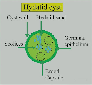
Hydatid Cyst


-Hydatid cysts are the larval stage of the tapeworm, Echinococcus granulosus.

-Dogs are the main hosts. Man and sheep are intermediate hosts.

-Hydatid disease is not uncommon amongst the mid-Wales farming communities, and up to 26% of farm dogs in this area have E. granulosus.

-The incidence is also high in some regions of the Mediterranean, North Africa, the Middle East, and Australia.

-If the ova are ingested by man, embryos are released when the chitinous coat is digested.

-These enter the liver by the portal vein. They may be destroyed, or they may develop into a cyst.

-In man, the cysts are found in the liver (65%), lung (25%), muscles (5%), bone (3%), and brain (1%). Each cyst is two-layered and contains straw-colored fluid in which there are free scolices, brood capsules containing scolices, and daughter cysts.

-Around the cyst is an area of compressed host tissue and fibrosis known as the pericyst. In 5–10% of cases, the cyst will die, and calcification may occur.

Preoperative Findings:

1. Hepatic cysts occur most frequently in the right lobe. A bacterial infection may result in a liver abscess. Rupture into a bile duct, or bile duct obstruction can occur and produce biliary colic. There may be jaundice. The number and location of the cysts are shown on a CT scan or ultrasound.

2. Pulmonary cysts can present with hemoptysis, dyspnea, cough, or chest pain. Chest X-ray may show a variety of appearances including an oval opacity, evidence of bronchial fistula formation, or rupture of the cyst with the development of a fluid level.

3. Eosinophilia occurs in about 30% of cases. The Casoni skin test is still used for screening. Immunoelectrophoresis is the most specific test. The complement fixation test is positive in up to 80% of cases. The hemagglutination test detects a specific antibody.

Anesthetic Problems:

1. Pulmonary hydatid cysts can cause bronchial obstruction, and occasionally they may rupture into the airway. If this happens, flooding of the lungs occurs, with widespread dissemination of the scolices.

2. Hydatid fluid is highly antigenic, and the rupture of a cyst has occasionally produced sudden death from an anaphylactic reaction. Anaphylaxis to hydatid was confirmed by increased serum histamine and tryptase levels, with elevated levels of total and echinococcus-specific IgE antibodies.

3. Patients can develop anaphylaxis of unknown origin as interventricular hydatid cystic mass.

4. Cerebral cysts can cause increased intracranial pressure.

5. Scolicidal agents are potentially toxic and their use in combination with surgery may increase the complication rate.

6. Postoperative complications following surgery for hydatid cyst of the lung included prolonged air leak and aspiration pneumonia.

Anesthetic Management:

1. Surgical removal is indicated, except in older patients with small cysts. Meticulous care must be taken to avoid the rupture and spread of the fertile scolices.

2. Relatively new drugs, such as mebendazole and albendazole, are being tested as scolicidal agents. However, there is no evidence that they are effective in the treatment of pulmonary hydatid.

3. Pulmonary cysts. Protective formalin-soaked packs are placed around the wound, an incision is made through the pericyst, and the cyst is carefully extruded by the anesthetist, using gentle hand ventilation.

4. Because of the risk of an anaphylactic reaction, epinephrine (adrenaline), metaraminol, isoprenaline, and steroids must be immediately available.

Budd–Chiari Syndrome

 Budd–Chiari Syndrome

Budd–Chiari Syndrome


-A syndrome caused by obstruction to the hepatic venous outflow and resulting in a clinical picture of hepatomegaly and portal hypertension. It may be secondary to hematological disorders, malignancy, oral contraceptives, heart failure, or constrictive pericarditis.

-The three main sites of obstruction are:

1-Inferior vena cava

2-Large hepatic veins

3-Small intrahepatic venules.

-The condition may be acute or chronic. Acute Budd–Chiari syndrome with hepatocyte necrosis may require urgent portosystemic, decompressive, surgical, or interventional radiological procedures.

-Untreated hepatic venous thrombosis usually results in progressive liver failure and death. Medical treatment is of little help, therefore shunt procedures or liver transplantation will be needed.

Preoperative Findings:

1. Abdominal pain and swelling are the commonest symptoms. Ascites and hepatomegaly will be present in the majority. Vomiting, splenomegaly, jaundice, or bleeding from esophageal varices may also occur. If the vena cava is involved there will be dependent edema.

2. Liver function abnormalities depend on the site and severity of the obstruction. A liver biopsy may show outflow obstruction, hepatic necrosis, and fibrosis.

3. Etiological factors include polycythemia, paroxysmal nocturnal hemoglobinuria, antiphospholipid syndrome, protein C deficiency, factor V Leiden, myeloproliferative disorders, and mechanical factors, such as webs and tumors. Hepatic venous thrombosis has been reported in association with ulcerative colitis, but in the patient described, iron deficiency had concealed an underlying polycythemia vera.

4. Treatment is aimed at the preservation of liver function and includes thrombolysis, angioplasty, stent placement, portacaval shunt, mesoatrial shunt, and liver transplantation.

Anesthetic Problems:

1. Hepatic function may be compromised.

2. Surgery may be required for portacaval or mesoatrial shunt or liver transplantation. This has been described in a patient with paroxysmal nocturnal hemoglobinuria.

3. Patients may present during pregnancy when the condition must be distinguished from HELLP syndrome and acute fatty necrosis of the liver. Antiphospholipid syndrome and preeclampsia in a primipara presented with Budd–Chiari syndrome from thrombosis of the right hepatic veins.

Thrombotic thrombocytopenic purpura resulted in postpartum hepatic venous thrombosis. Paroxysmal nocturnal hemoglobinuria has been associated with hemolytic crises and Budd–Chiari syndrome during pregnancy. Maternal deaths have been reported.

Anesthetic Management:

1. Hematological examination is important because, in the absence of mechanical causes for hepatic vein thrombosis, there is a high incidence of underlying hematological abnormalities; these include myeloproliferative disorders, and paroxysmal nocturnal hemoglobinuria, systemic lupus erythematosus, and antithrombin III deficiency.

2. Assessment of liver function, including coagulation.

3. Low-dose heparin infusion from the first postoperative day has been recommended, with subsequent low-dose aspirin and long-term anticoagulation to reduce the risks of graft thrombosis.

4. Liver transplantation will cure the inherited thrombophilias, for example, factor V Leiden, protein C and S, etc.

5. In a patient with paroxysmal nocturnal hemoglobinuria who developed Budd–Chiari syndrome, the resolution was achieved following bone marrow transplantation.

6. Pregnant patients with paroxysmal nocturnal hemoglobinuria should be carefully monitored for the onset of Budd–Chiari syndrome.

Esophageal Achalasia

Esophageal Achalasia

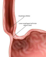
Esophageal Achalasia

Definition:

A chronic, progressive motor disorder of the esophagus associated with degenerative changes in the myenteric ganglia and vagal nuclei.

Components:

There are three components:

1-Failure of the lower esophageal sphincter to relax, with an increased resting sphincter pressure, which together results in a functional obstruction

2-Absence of sequential peristalsis in response to a bolus of food

3-Dilated, contorted esophagus

Pathophysiology and Management:

-Degeneration of the myenteric plexus and decreased nitric oxide synthesis may be the problem.

-Overspill may produce bronchopulmonary complications, and 5–10% of patients ultimately develop carcinoma of the esophagus.

-Nitrates and calcium channel blockers given before meals sometimes produce symptomatic improvement, but the mainstays of treatment are esophageal dilatation and surgical myotomy.

-Open surgery has been mostly replaced by laparoscopic myotomy and fundoplication.

-For elderly patients, an endoscopic injection of botulinum toxin can give relief for several months without the risk of surgery.

Preoperative Findings:

1. Symptoms include; dysphagia, retrosternal pain, regurgitation, and weight loss. In young people, the condition may be misdiagnosed as anorexia nervosa or asthma.

2. Respiratory complications, which may be attributed to asthma or chronic bronchitis, are secondary to the overspill of undigested material.

3. Nocturnal coughing occurs in 30%, and bronchopulmonary complications in 10% of patients.

4. The aspiration of larger volumes may result in lobar collapse, bronchiectasis, or lung abscess.

5. Rarely, it may present with a cervical mass and acute upper respiratory tract obstruction, necessitating urgent intervention.

6. There is an increased risk of esophageal carcinoma.

7. Diagnosis can be made on barium swallow, manometric studies, and endoscopy. Occasionally, acute dilatation may be seen on CXR, in which case, abnormal flow–volume curves will indicate variable intrathoracic tracheal obstruction.

Anesthetic Problems:

1. A predisposition to regurgitation and pulmonary aspiration in the perioperative period.

2. Passage of the tracheal tube past the dilated esophagus can be achieved with difficulty.

3. During recovery from anesthesia, neck swelling, and venous engorgement can be precipitated by coughing or straining. Acute thoracic inlet obstruction with stridor, deep cyanosis of the face, and hypotension can occur.

4. Upper airway obstruction or respiratory failure, particularly in the elderly. Rarely, an acute dilatation of the esophagus results in total airway obstruction.

5. Acute respiratory failure can occur after surgery.

6. The opening pressure of the cricopharyngeus muscle from above is much lower than that from below, therefore progressive dilatation of the upper esophagus may occur, particularly in association with mask ventilation or IPPV.

7. An increased intrathoracic pressure produced by a Valsalva maneuver forces air from the thoracic into the cervical esophagus. Occasionally, death can occur.

8. If acute airway obstruction is present, sudden decompression of the esophagus may cause the pharynx to flood with food and fluid, resulting in aspiration.

Anesthetic Management:

1. If anesthesia is required, precautions must be taken to reduce the risk of aspiration of gastric contents. The dilated esophagus must be emptied and decompressed. This needs a period of prolonged starvation, possibly with washouts of the esophagus.

2. A rapid sequence induction should be undertaken with awake tracheal extubation, the patient should be nursed in the lateral position during recovery.

3. Sublingual nifedipine 10–20 mg has been shown to reduce the basal sphincter pressure after 10 min and the effect lasts for up to 40 min.

4. Management of acute upper airway obstruction secondary to tracheal compression has been reported using the following methods:

a) Sublingual glyceryl nitrate.

b) Passage of a naso-esophageal tube.

c) Transcutaneous needle puncture.

d) Tracheal intubation.

e) Rigid esophagoscopy.

f ) Emergency tracheostomy.

g) Cricopharyngeus myotomy.

5. Treatment can be either surgical or medical. For the elderly and less fit patients, pneumatic dilatation, or endoscopic injection of botulinum toxin, may be appropriate. Heller myotomy and partial fundoplication can be performed either as an open or a laparoscopic procedure.