Educational Blog about Anesthesia, Intensive care and Pain management

Showing posts with label Regional anesthesia. Show all posts

Ultrasound Artifacts

Ultrasound Artifacts


Ultrasound Artifacts


1-Reverberation artifact:

➧ The processing unit in the ultrasound machine assumes echoes return directly to the processor from the point of reflection. 

➧ Depth is calculated as D = V × T, where V is the speed of sound in biological tissue and is assumed to be 1,540 m/sec, and T is time. 

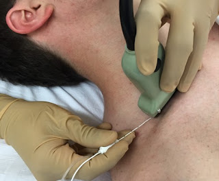
➧ In a reverberation artifact, the ultrasound waves bounce back and forth between two interfaces (the lumen of the needle) before returning to the transducer. 

➧ Since velocity is assumed to be constant at 1,540 m/sec by the processor, the delay in the return of these echoes is interpreted as another structure deep into the needle and hence the multiple hyperechoic lines beneath the block needle (Figure 1). 

Ultrasound Artifacts
Figure 1: Reverberation artifact


2-Mirror artifact:

➧ A mirror artifact is a type of reverberation artifact. 

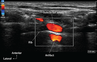
➧ The ultrasound waves bounce back and forth in the lumen of a large vessel (subclavian artery). 

➧ The delay in the time of returning waves to the processor is interpreted by the machine as another vessel distal to the actual vessel (Figure 2). 

Ultrasound Artifacts
Figure 2: Mirror artifact


3-Bayonet artifact:

➧ The processor assumes that the ultrasound waves travel at 1,540 m/sec through biological tissue. However, we know that there are slight differences in the speed of ultrasound through different biological tissues. 

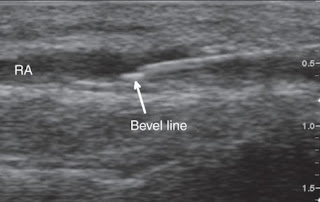
➧ The delay in the return of echoes from tissue that has a slower transmission speed, coupled with the processor’s assumption that the speed of ultrasound is constant, causes the processor to interpret these later returning echoes from the tip of the needle traveling in tissue with slower transmission speed as being from a deeper structure and thus giving a bayoneted appearance. 

➧ If the tip is traveling through tissue that has a faster transmission speed, then the bayoneted portion will appear closer to the transducer (Figure 3). 

Ultrasound Artifacts
Figure 3: Bayonet artifact


4-Acoustic Enhancement artifact:

➧ Acoustic enhancement artifacts occur distal to areas where ultrasound waves have traveled through a medium that is a weak attenuator, such as a large blood vessel. 

➧ Enhancement artifacts are typically seen distal to the femoral and the axillary artery (Figure 4). 

Ultrasound Artifacts
Figure 4: Acoustic enhancement artifact


5-Acoustic shadowing:

➧ Tissues with high attenuation coefficients, such as bone, do not allow the passage of ultrasound waves. 

➧ Therefore any structure lying behind tissue with a high attenuation coefficient cannot be imaged and will be seen as an anechoic region. (Figure 5). 

Ultrasound Artifacts
Figure 5: Acoustic shadowing artifact


6-Absent blood flow:

➧The Color-Flow Doppler may not detect blood flow when the ultrasound probe is perpendicular to the direction of blood flow (Figure 6). 

➧ A small tilt of the probe away from the perpendicular should visualize the blood flow (Figure 7, Figure 8). 

➧ Alternatively, for deep vascular structures, signals may be lost due to attenuation. 

➧ Increasing gain, while in Doppler Color-Flow mode, will increase the intensity of the returning signals, which may detect blood flow that was not previously detected. 

Ultrasound Artifacts
Figure 6: Radial a. Absent blood flow artifact
Ultrasound Artifacts
Figure 7: Radial a. Probe tilted away from the direction of blood flow
Ultrasound Artifacts
Figure 8: Radial a. Probe tilted towards the direction of blood flow

Accidental Total Spinal Anesthesia

Accidental Total Spinal Anesthesia


Accidental Total Spinal Anesthesia

Definition:

➧ A syndrome of the central neurological blockade. 

➧ It occurs when a volume of local anesthetic (LA) solution, intended for epidural anesthesia, enters the subarachnoid space and ascends to the cervical region.

Causes: 

a) After a known dural tap: 

➧ Accidental total spinal analgesia may occur in association with the original epidural after a known dural tap. 

b) After a top-up dose: 

➧ Following a top-up dose, as a result of accidental puncture of the dura by the epidural catheter. 

➧ High spinal anesthesia has occurred after top-ups of epidural catheters. 

➧ This is unlikely to result from catheter migration but may happen as a result of accidental puncture of the dura by the epidural catheter. 

➧ If part of the catheter lies within the epidural space and part within the subarachnoid, with a slow injection of LA, the solution will emerge from the proximal holes, and with a rapid one from the more distal. 

Presentation: 

1-Unexpected rapid rise in the sensory level. 

2-Numbness or weakness in the upper extremities. 

3-There is severe hypotension and bradycardia secondary to blockade of the sympathetic outflow. Occasionally cardiac arrest occurs. 

4-Rapidly increasing paralysis involves the respiratory muscles, due to phrenic nerve paralysis, resulting in apnea and hypoxia. 

5-The pupils become dilated and consciousness is lost. 

6-Cardiovascular collapse usually takes place immediately after the injection, although delays of up to 45 min. have been reported. 

7-Deaths have occasionally been reported. 

Duration: 

➧ Apnea may vary from 20 min. to 6 h, unconsciousness from 25 min. to 4 h, while full recovery of sensation may take up to 9 h. 

➧ The lengths of time vary with the agent, the dose, and the volume of LA given. Bupivacaine lasts longer than lidocaine. 

Management: 

a) Precautions: 

1-A test dose of LA is recommended. 

2-The injection of 3 ml of LA containing epinephrine 1:200 000, followed by an adequate pause to assess the effects, has been suggested. 

3-It has been recommended that, if a dural puncture occurs during active labor when a cesarean section is required, then further attempts should not be made. Either a spinal or a general anesthetic should be employed as an alternative. 

b) If Total Spinal a. occurs: 

1-Call for help. 

2-A non-pregnant patient should be turned supine, and the legs elevated to encourage venous return. The pregnant patient should be tilted in the lateral position to prevent aortocaval compression

3-The lungs should be inflated with oxygen

4-A tracheal tube can then be inserted. IPPV may have to be continued for up to 2 h, depending upon the LA and the volume used

5-Intravenous fluids should be infused rapidly

6-A vasopressor agent such as Ephedrine IV in 5–10 mg increments up to 30 mg is recommended. Epinephrine (adrenaline) 0.1–0.5 mg may occasionally be required but should preferably be avoided in patients in labor

7-Atropine for bradycardia, use epinephrine if the patient is unstable

Failed Spinal Anesthesia

Failed Spinal Anesthesia

Introduction:

➧ Spinal (intrathecal) anesthesia is one of the most reliable regional block methods: the needle insertion technique is relatively straightforward, with cerebrospinal fluid (CSF) providing both a clear endpoint of successful needle placement and a medium for carriage of local anesthetic (LA) within subarachnoid space. However, the possibility of failure has long been recognized, with an incidence of less than 1% in experienced hands.

➧ Literally, the word failure implies that spinal anesthesia was attempted, but no block resulted or block results, but is inadequate for the proposed surgery. 

➧ Such inadequate block may be related to the three components of the block: the extent, the quality, or the duration of local anesthetic action, often with more than one of these being inadequate.

➧ The intrathecal injection can go astray within each of the five phases of an individual spinal anesthetic, leading to blocking failure, these being, in sequence: 

1-Lumbar puncture 

2-Solution injection 

3-Spreading of the drug through CSF 

4-Drug action on the spinal nerve roots and cord 

5-Subsequent patient management

Causes and prevention of failure:

1-Unsuccessful Lumbar Puncture (Dry Tap):

-Inability to obtain CSF ‘Dry Tap’, is the only cause of failure which is immediately obvious. 

Causes:

a) Incorrect needle insertion or Poor patient positioning:

Prevention: 

➧ A calm, relaxed patient is more likely to assume and maintain the correct position, so: 

-Explanation (before and during the procedure) and gentle, unhurried patient handling are vital.

-Premedication with light anxiolytic for relaxing the patient. 

-Local anesthetic infiltration at the puncture site is effective without obscuring the landmarks.

-Systemic analgesia (IV or inhalation) helps in achieving the correct position for patients in pain (e.g. from a fractured hip).

b) Anatomical abnormalities of the spine:

-Kyphosis, scoliosis, calcification of ligaments, consequences of osteoporosis, obesity, and patient anxiety, make both positioning the patient and needle insertion more difficult, especially in the elderly. 

Prevention: 

-Good clinical training is the key to success.

-Adherence to the basic rules of positioning, needle insertion, and use of adjuncts.

-Lateral or paramedian approach, especially if the mid-line ligaments are heavily calcified. 

-Ultrasound guidance: a pre-procedure scan can be useful in patients with anatomical abnormality to identify the midline and level of injection and to assess the depth of dura from the skin.

c) Equipment-related factors: A blocked needle lumen:

Prevention: 

-Both needle and stylet must be checked for correct fitness before use.

-The needle should not be advanced without the stylet in place, because tissue or blood clots can easily obstruct the fine-bore needles used now.

-Prompt needle withdrawal and 'flush test' to assure patency.

2-Pseudo-successful Lumbar Puncture:

-The appearance of clear fluid at the needle hub is usually the final confirmation that the subarachnoid space has been entered.

a) Epidural 'Top-up' dose:

-Rarely, the clear fluid is not CSF, but LA injected as an epidural ‘top-up’ dose or spreading from the lumbar plexus.

Prevention:

-Unfortunately, a positive test for glucose in the fluid does not confirm that this fluid is definitely CSF because extracellular fluid constituents diffuse rapidly into fluids injected into the epidural space.

b) Congenital arachnoid cyst:

-Another, rarer cause is a congenital arachnoid cyst (Tarlov cyst), which are meningeal dilatations of the posterior spinal nerve root, present in 4.5-9% of the population.

3-Solution Injection Errors:

1. Dose selection:

-The dose injected, within the normal range, has only a small effect on the height of a spinal block but is important in determining the quality and duration of the block. 

➧ The dose chosen will depend on: 

-The specific LA used

-The baricity of LA solution

-The patient’s subsequent posture

-The type of block intended

-The extent and duration of planned surgery

Causes: 

➧ Some anesthetists use lower doses than is traditional, in attempts to either: 

-Minimize hypotension, by producing a unilateral block. 

-Decrease block duration which speeds postoperative mobilization and decreases the need for bladder catheterization.

➧ Such lower doses will increase the margin for error and exaggerate the consequences of other problems such as: 

-Loss of injectate and so risk an inadequate block.

-The ‘dead space’ of the needle and hub will contain a significant proportion of what is a small volume to start with.

2. Loss of injectate:

Causes: 

-Leakage of LA solution through the Luer connection between syringe and needle. 

-Leakage through a defect at the junction of needle hub and shaft. 

➧ Given the small volumes involved, the loss of a few drops can cause a significant decrease in the mass of the drug reaching the CSF, and thus in its effectiveness. 

Prevention: 

-Insert the syringe containing the injectate firmly into the hub of the needle, and check that no leakage occurs.

3. Misplaced injection:

Causes: 

➧ Anterior or posterior displacement of the needle tip from subarachnoid to epidural space:

a) During connection of the syringe to the needle, where deposition of a spinal dose of LA will have little or no effect. 

b) During fluid aspiration for confirmation that the needle tip is still in the correct space, may displace the tip unless performed carefully, as may the force of the injection of the syringe contents. 

Prevention:

-The dorsum of one hand should be anchored firmly against the patient’s back and the fingers are used to immobilize the needle, while the other hand is used to manipulate the syringe.

-After attachment of the syringe, aspirate 0.5-1 ml to confirm the free flow of CSF, and at the end of the spinal injection, aspirate 0.5-1 ml, to confirm that the needle tip is still in the subarachnoid space, the aspirated volume is re-injected before the needle is withdrawn. Some anesthetists advocate that this is done halfway through as well, 

c) Tip displacement is an important issue with the ‘Pencil point’ needles, as the opening at the end of these needles is proximal to the tip, so only a minor degree of ‘backward’ movement during syringe attachment may result in epidural injection. 

-Also, the opening of these needles may ‘straddle’ the dura so that some solution reaches the CSF and some of the epidural space. 

-This may be exaggerated by the dura acting as a ‘flap’ valve across the needle opening. Initially, CSF pressure pushes the dura outwards so that aspiration is successful, but subsequent injection pushes the dura forward and the solution is misplaced. 

-A variant is that the needle tip penetrates the dura, but it is the arachnoid mater that acts as the flap valve so that accidental subdural injection results. 

Prevention: 

-Rotation of the needle through 360 degrees after the initial appearance of CSF, and before check aspiration, as the rotation reduces the risk of the membrane edges catching on the opening.

4. Inadequate intrathecal spread:

➧ Factors affecting the intrathecal spread of a local anesthetic solution: 

-Anatomy of the vertebral canal.

-Solution physical characteristics.

-Gravity.

a) Anatomical abnormality: 

-Abnormalities of the curves of the vertebral column as kyphosis or scoliosis, may interfere with the solution spread.

Examination of the patient should reveal whether this might occur, but it is not possible to predict whether the effect will be excessive spread or failure. 

-A rare possibility, is that the ligaments supporting the spinal cord within the theca, form complete septae which act as longitudinal or transverse barriers to LA spread. This can result in a block that is entirely unilateral or limited cephalad spread.

-Spinal stenosis or other pathological lesions can limit the spread, effectiveness, or both.

-Previous spinal surgery or intrathecal chemotherapy may result in adhesions that interfere with LA spread. 

-Increased CSF volume in the lumbar theca can cause restricted cephalad spread of intrathecal injection.

-A variation of this factor is dural ectasia, which is a pathological enlargement of the dura seen in most patients with Marfan’s syndrome and in some other connective tissue disorders. 

b) Solution density (baricity):

-Isobaric solutions, with a density within the normal range of CSF, will block the lower limbs with little risk of thoracic nerve block and thus less hypotension. 

-Plain solutions of bupivacaine, although referred to as isobaric, are actually of lower density to be hypobaric at body temperature (37ºC). They have a less predictable spread than that of a truly isobaric preparation, and the block may be not higher than the second lumbar dermatome with slow onset. 

-Hyperbaric solutions, with a density greater than that of CSF, move under the combined influence of gravity and the curves of the vertebral canal. If the patient is placed supine after the injection of a hyperbaric preparation at the mid-lumbar level, the solution will spread ‘down’ the slope under the effect of gravity to pool at the ‘lowest’ point of the thoracic curve, so exposing all nerve roots up to that level to an effective concentration of LA. (Figure 1)

-However, if a lumbar puncture is performed at the fourth lumbar or the lumbosacral interspace, LA may be ‘trapped’ below the lumbar curve, especially if the patient is in the sitting position during injection and maintained in that position for a period thereafter. This results in a block that is restricted to the sacral segments.

Effects of drug baricity
Figure 1: Effects of drug baricity

Prevention:

-Avoiding too low injection level unless a deliberate ‘saddle’ block is intended.

4. Ineffective drug action:

➧ The solution injected reaches the target nerves, but it is ineffective or inactive, with a variety of possibilities: 

a) Incorrect drug injection (Identification error): 

-Using LA for skin infiltration or analgesic adjuvants, used from the same sterile preparation area, instead of spinal LA, may lead to an ineffective block. 

Prevention:

-The use of labeled syringes, but this is not easy within a sterile field. 

-The use of syringes with different sizes for each component of the procedure.

-Minimizing the number of ampoules on the block tray (such as using the same LA for both skin infiltration and spinal anesthesia).

b) Physico-chemical incompatibility: 

-The mixing of two different pharmaceutical preparations raises the possibility of ineffectiveness as a result of the interaction between LA and adjuvant.

-Chemical reaction can generate an obvious precipitate, or lower the pH of the LA solution which will decrease the concentration of the unionized fraction which diffuses into nerve tissue resulting in a decreased effect.

-Local anesthetics are compatible with most opioids, but the situation is less definitive with other adjuvants such as clonidine, midazolam, ketamine, and other substances. 

-The stability is unknown when mixing three or more substances together for intrathecal use. 

-The incidence of failure is greater after the addition of a vasoconstrictor solution. 

c) Inactive LA solution: 

-Ester-type LAs, are chemically labile so that heat sterilization and prolonged storage, particularly an aqueous solution, can make them ineffective because of hydrolysis, and hence they need very careful handling. 

-Amide-type LAs (e.g. lidocaine, bupivacaine, etc.) are more stable and can be heat sterilized and stored for several years without loss of potency. 

d) Local anesthetic resistance:

-Very rarely, failed spinal block has been attributed to physiological resistance to the actions of LA drugs.

-This problem is due to the mutation of sodium channels (channelopathy) which is associated with significant neurological diseases such as; intractable epilepsy and chronic pain, however, this does not exist in asymptomatic individuals.

-A history of repeated failure of dental or other LA techniques is accompanied by speculation that the problem is due to sodium channel mutation that renders the drugs ineffective. 
4-Failure of Subsequent management:

➧ Not all of a patient’s claims of discomfort, or pain, during spinal anesthesia, are due to inadequate block.

Causes:

-Lying awake during surgery is not a pleasant experience for most patients, and anxiety alone can cause patient discomfort. 

-Furthermore, operating tables are designed for surgical access, not patient comfort; and intra-abdominal stimuli can result in afferent impulses in unblocked parasympathetic nerve fibers causing unpleasant sensations. 

Prevention: 

-Good preoperative patient counseling followed by a supportive approach from the anesthetist during the operation is important in avoiding such problems. 

-Judicious, and proactive use of systemic analgesic drugs. 

-Sufficient sedation to produce drowsiness, or even sleep (with appropriate monitoring), is indicated except in obstetric situations, where small doses may be useful.

-Distraction techniques such as listening to music.

5-Testing the block:

-It is mandatory to test the level of the block before surgery commences

-Most patients will have some anxiety about the effectiveness of the injection, and this will be increased if testing is started too soon. 

-Conventional practice is to check motor block by testing the ability to lift the legs, followed by testing of sensory block to stimuli such as light touch, cold, or pin-prick.

-It is advisable to start testing in the lower segments, where onset will be fastest, and work upwards. Proving early that there is some effect encourages patient confidence; testing too soon does the opposite. 

-Establishing that the level of block is appropriate for the projected surgery is often taken to demonstrate that the quality of block is adequate also. 

-A covert pinch of the site of the proposed surgical incision may be a better indicator of skin analgesia and can be reassuring if the block has been slow in onset. Asking the surgeon to do the same with toothed surgical forceps after distracting the patient with conversation.

6-Catheter and Combined Techniques:

-The majority of spinal anesthetics involve a single shot of LAs. 

-To take advantage of the rapid onset and profound block of spinal anesthesia, both continuous and combined spinal-epidural techniques have been introduced to increase flexibility. 

-If the catheters are correctly placed, problems of inadequate spread, quality, and duration of effect can be dealt with. 

-However, insertion of an intrathecal catheter can be difficult to achieve in some patients and can result in the misdirection of the LA solution, with the risk of neurotoxicity.


Read more: ☛ Failed Epidural Block

Management of Failed Spinal Anesthesia

Management of Failed Spinal Anesthesia


Management of Failed Spinal Anesthesia
➧ Failure of a spinal anesthetic is an event of significant concern for both patient and anesthetist when it is immediately apparent, but it can have serious consequences (clinical and medico-legal) if the problem only becomes evident once surgery has started.

➧ This can be a source of pain, anxiety, and psychological trauma to the patient and a cause of stress, complaints, and medico-legal sequelae to the anesthetist.

Prevention is better than cure:

➧ The spinal block should be performed with meticulous attention to detail. 

➧ If there is any doubt about the nature or duration of the proposed surgery, a method other than standard spinal anesthesia should be used.

Management of failed spinal block:

-The precise management of a failed spinal block will depend on the nature of the inadequacy and the time at which it becomes apparent. 

-The slower the onset of either motor or sensory block, the more likely is the block to be inadequate, so the more detailed assessment should be. 

-While the onset of spinal anesthesia is rapid in most patients, it can be slow in some; so, ‘transient time’ should always be allowed. 

-However, if the expected block has not developed within 15 min., some additional maneuver is needed, as follows:

1-No block:

Causes:

-Incorrect or ineffective solution was injected. 

-Solution has been deposited in the wrong place. 

Management: 

-Repeating the block or conversion to general anesthesia is the only option.

2-Spinal block of inadequate height:

Causes:

-Some injectate has been lost or misplaced. 

-The level of injection was too low.

-Anatomical abnormality has restricted spread.

Management:

-If a hyperbaric solution was used, flex the patient’s hips and knees and tilt the table head down (Trendelenburg position). This straightens out the lumbar lordosis but maintains a cephalad ‘slope’ and allows any solution ‘trapped’ in the sacrum to spread further. 

-In an obstetric situation, turn the patient to the full lateral position with a head-down tilt and reverse the side after 2–3 min.

-If a plain (and usually slightly hypobaric) solution has been used, it may help to sit the patient up but beware of peripheral pooling of blood.

-If an intrathecal catheter injection results in an inadequate spread, do not inject more of the same solution because the dose has minimal effect on the intrathecal spread. 

-Either posture should be manipulated as above, or a different baricity of solution should be tried, or the catheter should be withdrawn before the injection is repeated.

3-Unilateral block:

Causes: 

-This is most likely because of positioning. 

-The longitudinal ligaments supporting the cord have blocked spread. 

Management:

-If the operation is on the anesthetized limb, the surgeon should know that the other leg has a sensation, and the patient should be reassured and closely monitored. 

-Otherwise, turning the patient onto the unblocked side if a hyperbaric solution was used (or the reverse for plain solutions) may facilitate spread.

4-Patchy block:

The block appears adequate in extent, but the sensory and motor effects are incomplete. 

Causes: 

-The local anesthetic (LA) dose was inadequate.

-The LA was partially misplaced.

Management:

-If this becomes apparent before surgery starts, the options are to repeat the spinal injection or to use IV analgesia, the latter being the only option after skin incision.

-It may not be necessary to recourse to general anesthesia, as sedation or analgesic drugs are often sufficient especially when patient anxiety is a major factor. 

-Infiltration of the wound and other tissues with LA by the surgeon may also be useful in such situations.

5-Inadequate duration:

Causes:

-An inadequate dose of LA was delivered to the CSF. 

-Syringe swap; Lidocaine (intended for skin infiltration) was confused for bupivacaine. 

-The operation has taken longer than expected.

Management: 

-Sedation, IV analgesia, or infiltration of LA may be adequate, but often the only option is to convert to general anesthesia.

Repeating the spinal block:

➧ If no effect at all was seen 15-20 min. following the injection, it seems reasonable to repeat the block, paying close attention to avoiding the potential pitfalls. 

➧ In all other situations besides total failure, there must be some LA already in the CSF, and anxieties relating to several issues have to be taken into account: 

1-A restricted block may be due to an anatomical factor, impeding the physical spread of the solution, and it may have the same impact on a second injection, resulting in a high concentration of LA at or close to the site of injection leading to neurotoxicity.
2-Barriers to spread within the subarachnoid space may also affect epidural spread (and vice versa), so an attempt at epidural block may not succeed either.

3-Repeated injection in response to a poor quality block may lead to excessive cephalad spread with the potential for cardiovascular instability, respiratory embarrassment, or total spinal anesthesia, so a lower dose should be used to reduce this risk. 

4-A good quality, but unilateral block, might lead to an attempt to place a second injection into the ‘other’ side of the theca, but the risk of placing the second dose on the same side must be significant. 

5-A block of inadequate cephalad spread might be overcome by repeating the injection at a higher level, but should only be attempted when there is a considerable indication for a regional technique.

6-When a repeat block is considered, the adjacent nerve tissue is already affected by LA action, so the risk of direct needle trauma is increased.

Recourse to general anesthesia:

➧ There are many ways in which an inadequate block might be ‘rescued’: 

-General anesthesia must be considered if one or two simple measures have not rectified matters. 

-Common sense and clinical experience are usually the best indicators of exactly when to convert to general anesthesia. 

-If general anesthesia is induced to supplement partially effective spinal anesthesia, any degree of sympathetic nerve block will make hypotension more likely.

Postoperative Management:

1-Documentation and follow-up:

-The anesthetic complication details should be fully documented in the notes. 

-The patient should be provided with an apology and a full explanation after the operation. 

-Giving the patient a written summary of events for presentation to a future anesthetist can be very helpful, although care should be taken to prevent medico-legal recourse.

-Rarely, the inadequate spread has been the first indication of pathology within the vertebral canal, so if there is any suspicion, look for symptoms and signs of neurological disease, and consult a neurologist. 

-During follow-up of a patient in whom no block was obtained, the possibility of LA ‘resistance’ may seem an attractive explanation.

2-Investigating LA effectiveness:

-Performing skin infiltration with some of the solutions intended for the spinal injection should demonstrate that it is effective. 

-If the concern continues the operating theater, pharmacy, and anesthetic department records should be cross-checked to see whether other practitioners in the hospital have experienced any problems. 

-Similarly, distributors should be able to check whether other hospitals, that have been supplied with material from the same batch, have reported difficulty.


Needle Misplacement during Caudal block

Needle Misplacement during Caudal block

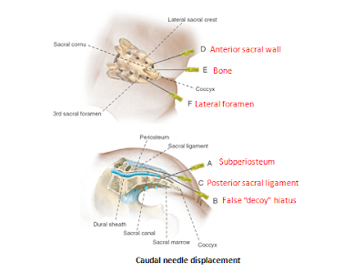
Sites of Misplacement:

A-Subperiosteal

B-False decoy hiatus

C-posterior sacral ligament

D-Anterior sacral wall

E-Bone

F-Lateral foramen


Needle Misplacement during Caudal block

Failed Epidural Block

Failed Epidural Block


Introduction:

➧ Failure of epidural anesthesia and analgesia occurs in up to 30% of clinical practices. 

➧ Some technical factors can help to increase the primary and secondary success rates.

A) Technical factors influencing block success:

1-Patient Position:

➧ Patient positioning potentially affects needle placement by changing the relationship between osseous and soft tissues. 

a) Sitting Position 

➧ Results in shorter insertion times and higher accuracy at the first attempt than in the lateral position. 

➧ Causes more vagal reflexes than lateral position. 

➧ This leads to epidural venous plexus distension, which may theoretically increase the risk of vascular puncture, especially in parturients. 

b) Lateral Position 

➧ Increases the distance from the skin to the epidural space. 

➧ Results in more technical difficulties compared with the sitting position.

2-Puncture Site:

➧ Inaccurate dermatomal block-level or anatomical landmarks of neuraxial puncture are not suitable for the type of surgery.

3-Approach:

a) Midline Approach 

➧ Results in a higher incidence of paresthesia than the paramedian approach. 

➧ Results in a bloody puncture in non-pregnant adults than paramedian approach. 

➧ The ligamentum flavum is not continuous in all patients, and the presence of midline gaps may make the loss of resistance (LoR) to needle advancement and injection of air/saline less perceptible when the midline approach is used. 

b) Paramedian Approach 

➧ Results in faster catheter insertion times, and less dependent upon spine flexion. 

➧ Paramedian catheters cause less epidural tenting and pass cephalad more reliably than midline catheters.

4-Identification of the Epidural Space:

➧ Correct placement obviously requires correct identification of the epidural space. The LoR using saline has become the most widely used method, while LoR to air and the hanging drop technique is less widely used with no difference in the success rate or adverse events, other than a 1.5% reduction in post-dural puncture headache [1] when using saline. 

➧ In obstetric epidurals, using saline for LoR results in fewer attempts than using air, but with comparable final success rates. 

➧ The use of the ‘preferred technique’ (i.e. the technique used by anesthetists 70% of the time) results in significantly fewer attempts, a lower incidence of paresthesia, and fewer dural punctures, irrespective of whether saline or air is used for LoR. 

➧ The hanging drop technique depends on negative pressure within the epidural space and is useful only in the sitting position. 

➧ Identification of the epidural space was reported at 2 mm deeper for the hanging drop when compared with LoR, possibly indicating an increased risk of dural puncture.

➧ Ultrasound pre-assessment of lumbar epidural space depth has been shown to correlate well with actual puncture depth in obese parturients. 

➧ The use of ultrasound led to less bony contact, a shorter time to block success, and decreased supplemental opioid requirements.

5-Epidural Catheter Location:

➧ Epidural catheters may primarily be placed incorrectly, or become dislodged during operation. 

➧ Primary misplacement of epidural catheters in the paravertebral space, in the pleural cavity, or intravascularly. 

➧ Transforaminal migration of the catheter tip and asymmetric spread during epidural analgesia. 

➧ During normal patient movement, epidural catheters may be displaced by centimeters. 

➧ Changes in epidural pressure and cerebrospinal fluid (CSF) oscillations can contribute to the displacement of epidural catheters. 

➧ Midline fat pedicles may form a barrier to the spread of local anesthetics (LA).

6-Catheter Insertion and Fixation:

➧ The catheter should be inserted at least 4 cm into the epidural space.

➧ Suturing of the epidural catheter is associated with less migration, but at the cost of increased inflammation at the puncture site. 

➧ Tunneling the epidural catheter for 5 cm is associated with less movement of the catheter and decreases catheter migration but it will not maintain the original position. 

➧ Tunneling caudal epidural catheter in children reduces the risk of bacterial colonization to levels comparable to un tunneled lumbar catheters because tunneling places the catheter entry point above the diaper in babies and toddlers. 

➧ For lumbar and epidural catheters, the advantages of tunneling are less obvious and the need to prevent dislodgement must be weighed against the increased incidence of erythema at the puncture site, potentially linked to increased risk of bacterial colonization.

➧ Catheter fixation devices are available which may significantly reduce migration percentage and reduce rates of analgesic failure.

7-Test Dose:

➧ A test dose is given with two main objectives of detecting intrathecal or intravascular catheter placement. 

➧ A test dose of lidocaine (to detect intrathecal placement) and epinephrine (to detect intravascular placement) is recommended in patients without contraindications to epinephrine. 

➧ Specific regimens to detect intravascular catheter position: 

-Fixed epinephrine test dose for non-pregnant adult patients. 

-Fentanyl test dose for parturients. 

-Weight-adjusted epinephrine test dose for children. 

➧ Patients sensitive to intravascular epinephrine (parturients, patients with cardiac or vascular disease) may experience undesirable side effects if the test is positive. However, this risk is outweighed by the systemic toxic effects of LA if the intravascular placement is not detected.

8-Equipments:

➧ The orifice of the catheter can lie laterally or anteriorly in the epidural space putting the LA more to one side and producing a unilateral block. In general, multi-orifice catheters are considered better than single-orifice catheters. 

➧ Manufacturing errors, such as faulty markings on the epidural catheter, can lead to a wrong depth of placement. 

➧ Debris in the catheter or disconnection can cause epidural failure. 

➧ Obstruction of the epidural infusion system by an airlock, as little as 0.3–0.7 ml of air, in the bacterial filter. 

➧ The knotting of the catheter internally or externally can cause obstruction. 

➧ Removal of a presumed knotted catheter can be attempted after sensation has returned to monitor neurological symptoms during catheter removal. When radicular symptoms or pain occur during the removal of a catheter, this should be immediately stopped. It has been found that removal is easiest if the patient is in the same position as at insertion. Surgical removal of a broken catheter is not compulsory if the patient remains asymptomatic.

References:


Read more: ☛ Failed Spinal Anesthesia

Local Anesthetic Toxicity

Local Anesthetic Toxicity


Local Anesthetic Toxicity

Clinical Manifestations & Management: 

1-CNS manifestations: 

➧ Early signs: circumoral numbness (Earliest sign), tongue paresthesia, dizziness

➧ CNS Excitation: (restlessness and agitation)

➧ CNS Depression: (slurred speech, drowsiness, unconsciousness)

➧ Muscle twitching, tonic-clonic seizures

➧ Respiratory arrest often follows

Management: 

1-Stop LA injection or infusion 

2-Call for help 

3-Oxygenation 

4-Hyperventilation (to decrease cerebral blood flow) 

5-Anti-seizures: Benzodiazepines (diazepam 0.1-0.2 mg/kg), Thiopental (1-2 mg/kg) 

2-Respiratory manifestations: 

➧ Local anesthetics (LAs) depress hypoxic drive (ventilatory response to low PaO₂)

➧ Apnea can result from phrenic and intercostal nerve paralysis

Management: 

➧ Respiratory support

3-CVS manifestations: 

➧ In general, LAs depress myocardial automaticity (spontaneous phase IV depolarization) and reduce the refractory period causing: bradycardia, heart block (varying degrees), and hypotension which may lead to cardiac arrest. 

➧ The R (+) isomer of bupivacaine rapidly blocks cardiac sodium channels and dissociates slowly. 

➧ Major cardiovascular toxicity requires about 3× the concentration required to produce seizures. 

➧ Ropivacaine is 70% less likely to cause severe cardiac arrhythmias than bupivacaine. 

Management: 

1-ACLS protocols 

2-Vasopressors: Ephedrine, Norepinephrine, Epinephrine

3-Lipid emulsion infusion: 

➧ Use of Lipid Emulsion: 

-IV bolus of Intralipid 20% (1.5 ml/kg over 1 min) about 100 ml and start infusion of (0.25 ml/kg/min) 

-If adequate circulation has not been restored: Repeat the bolus dose twice at 5 min intervals and increase the infusion rate to (0.5 ml/kg/min)

-Continue infusion until adequate circulation has been restored

-Measure LA & triglyceride levels

Patient-related factors to consider during large volume peripheral nerve block (PNB): 

1-Age: 

➧ Newborns have about half the adult concentration of α-acid glycoprotein (AAG) which binds free LAs. 

➧ Persons over 70 years show increased sensitivity to LAs and decreased clearance. 

Recommendation: Reduce LAs dose by 10-20%. 

2-Renal dysfunction: 

➧ There may be a change in clearance of LAs in uremic patients. 

➧ Uremic patients show a rapid rise in LAs plasma levels probably secondary to a hyperdynamic circulation, but uremic patients have increased AAG levels. 

Recommendation: Reduce LAs bolus and continuous dose by 10-20% in uremic patients. 

3-Hepatic dysfunction: 

➧ Clearance of LAs can be dramatically decreased but plasma concentrations remain close to normal secondary to increased volume of distribution (Vd). These patients also can have renal and cardiac dysfunction. 

Recommendation: Initial bolus dose can be in the normal dose range but the continuous infusion dose should be reduced by 10-50%. 

4-Heart Failure: 

➧ Decreased blood flow to the liver and kidneys can cause a decrease in clearance

Recommendation: Repeat or continuous dosing of LAs should be reduced by 10-20%

5-Pregnancy: 

➧ Progesterone may increase the sensitivity of nerve axons

➧ There is an enhanced risk of cardiotoxicity by bupivacaine and ropivacaine induced by progesterone. Increased cardiac output causes increased uptake of LAs

Recommendation: Avoid large volume PNB in 1st trimester and reduce doses in epidural and spinal anesthesia in pregnancy

6-Drug interaction: 

➧ Amide LAs are cleared by the liver cytochrome P450 enzymes

➧ Propanolol, Cimetidine and Itraconazole can decrease bupivacaine clearance by 30-35%

➧ Ciprofloxacin and Fluvoxamine decrease the clearance of ropivacaine

Recommendation: Single bolus dose is of little concern but continuous infusion should be altered (10-20% decrease)