Educational Blog about Anesthesia, Intensive care and Pain management

Complications while ETT in place

Complications while ETT is in place

A) Obstruction of the endotracheal tube (ETT):

Causes:

1-Accumulation of secretions in the tube 

2-Kinking of the tube.

B) Misplacement or migration of the ETT:

➧ A common complication and right mainstem intubation has been associated with increased mortality in critically ill patients. 

➧ Traditional methods of assuring proper tube position include: 

-Observing bilateral chest expansion.

-Auscultation of bilateral breath sounds and the lack of air in the epigastrium. 

➧ Unfortunately, none of these methods is reliable, leading to the use of capnography as the current gold standard to detect placement of the tube in the airway and rule out esophageal placement. 

➧ Capnography, however, does not detect proximal or distal placement of the ETT within the airway.

C) Respiratory Complications of TI:

1-Dead Space

➧ Tracheal intubation usually results in a small reduction in dead space ventilation (25-75 ml); this reduction is too small to be beneficial in most patients.

2-Airflow Resistance

➧ Resistance to airflow through ETT is significant, particularly at small tube diameters. 

➧ Most airflow through ETTs is turbulent. The resistance to flow is therefore proportional to the fifth power of the radius. This can result in marked differences in the pressure necessary to cause airflow depending on the internal diameter of the ETT and the flow rate required.

3-Airway Resistance

➧ Intubation also causes an increase in the lower airway resistance in most people as a result of parasympathetic activation of airway smooth muscle. Generally, the increase in lower airway resistance is not clinically significant.

4-Bronchospasm

➧ TI can trigger severe bronchospasm. Interestingly, only one-half of the patients in whom bronchospasm occurred had a previous history of bronchial asthma or pulmonary disease. 

➧ Preoperative inhaled β-agonists such as albuterol as well as anticholinergic agents may prevent bronchospasm in patients with reactive airway disease. 

➧ Inhaled/IV β-agonists, anticholinergic agents such as glycopyrrolate, and an increase in the level of inhaled anesthetic agents; all have been used intraoperatively to treat bronchospasm. 

➧ In some instances, the tracheal tube must be removed before the bronchospasm will stop.

5-Cough

➧ ETT results in reduced cough efficiency and, if humidification is not provided, drying of the upper airway.

6-PEEP

➧ The presence of ETT through the glottis reduces the small positive pressure or "auto-PEEP" caused by the resistance of air flowing through the glottis. This loss of PEEP may reduce the functional residual capacity in some patients in respiratory failure dependent on "auto-PEEP" for oxygenation.

7-Respiratory Mucosa

➧ The presence of an artificial airway leads to functional and morphologic changes in the respiratory mucosa due to the loss of the humidification, warming, and filtering effects of the upper airway (especially the nasopharynx). In the long term, this may result in squamous metaplasia and granulation tissue formation.


Hemodynamic effects of Laryngoscopy and TI

Hemodynamic effects of Laryngoscopy and TI


Autonomic innervation and response of the airway:

➧ The area of the trachea and pharynx is richly innervated and involves both the parasympathetic and sympathetic nervous systems.

➧ Following the mechanical stimulation of the upper respiratory tract (URT) (i.e. nose, epipharynx, laryngopharynx), the afferents are carried by the glossopharyngeal nerve and from the tracheobronchial tree via the vagus nerve which enhances the activities of the cervical sympathetic afferent fibers resulting in a transient rise in heart rate (HR) and blood pressure (BP). 

➧ The lower respiratory tract (LRT) is protected by reflex arcs from both the upper and lower airways. The afferent pathways are comprised of the glossopharyngeal nerves in the oropharynx, superior to the anterior surface of the glottis, and the superior and recurrent laryngeal nerves for the posterior and inferior glottis, whereas the efferent pathway is controlled by the vagus. 

➧ Afferent stimuli can therefore trigger cardiac, airway, cerebral, neuromuscular, and adrenal responses. 

➧ The hemodynamic responses to orotracheal intubation have two components. The first is the response to laryngoscopy and the second is the response to tracheal intubation (TI). 

➧ Tachycardia and hypertension have been reported since 1950 during intubation under light anesthesia as TI causes a reflex increase in sympathetic activity. 

➧ The hemodynamic responses are due to reflex sympathoadrenal discharge provoked by epilaryngeal and laryngotracheal stimulation after laryngoscopy and TI, this results in hypertension, tachycardia, arrhythmia, and a change in plasma catecholamine concentrations. 

➧ Translaryngeal intubation of the trachea stimulates laryngeal and tracheal receptors, resulting in a marked increase in the elaboration of sympathomimetic amines. This sympathetic stimulation results in tachycardia and a rise in BP. 

➧ In normotensive patients, this rise is approximately 20-25 mmHg; it is much greater in hypertensive patients. Nasopharyngeal intubation causes a significant pressor response. 

➧ Stimulation of the larynx and trachea by the passage of the tracheal tube, but not direct laryngoscopy, causes a significant increase in this response. Direct stimulation of the trachea appears to be a major cause of the hemodynamic changes associated with TI. 

➧ The extent of the reaction is affected by many factors: the technique of laryngoscopy and intubation, and the use of various airway instruments. Laryngoscopy itself is one of the most invasive stimuli during orotracheal intubation. 

➧ Many anesthesiologists agree that applying a small force to the patient’s larynx when using a laryngoscope might prevent excessive hyperdynamic responses to orotracheal intubation. 

➧ In infants, laryngoscopy and TI often result in bradycardia from vagal stimulation. Administration of anticholinergic agents such as atropine can block this response. In older children or adults, this vagal response is rarely observed. Although bradycardia can develop in up to 10% of patients undergoing TI, the typical result is tachycardia and hypertension, leading to an increase in myocardial oxygen consumption. It has been shown that up to 15% of patients undergoing TI under general anesthesia will have ventricular arrhythmias, with the majority of events occurring at the time of tube insertion, as opposed to at the time of laryngoscopy. 

➧ The rise in HR and BP occurs about 14 sec. after the start of direct laryngoscopy and becomes maximal after 30-45 sec. Prolonged intubation time in difficult airways, in addition, induces hypercarbia and decreases anesthetic gas concentration, resulting in tachycardia and hypertension. These responses are usually transient and innocuous.

Cardiac Patients:

➧ In patients with co-existing hypertension or ischemic heart disease, these may be exaggerated or may jeopardize the balance between myocardial oxygen requirements and delivery. In these patients, it is important to minimize the duration of direct laryngoscopy, if possible, to less than 15 sec. 

➧ During and immediately following TI associated with tachycardia and hypertension, there is a decrease in the left ventricular ejection fraction (stroke volume/end-diastolic volume). This is particularly marked in patients with coronary artery disease. 

➧ Increases in HR may be associated with ST-segment changes that indicate myocardial ischemia. 

➧ The cardiovascular response to TI can be problematic if the patient suffers from cardiac disease, cerebrovascular or abdominal-vascular disease in which hypertension may lead to hemorrhage.

Hypertensive Patients:

➧ Hypertensive patients are prone to greater and exaggerated circulatory responses after laryngoscopy and TI, because of long-term persistent vascular hyperreactivity, than normotensive patients. An increase in BP associated with TI is dangerous and may cause complications, including pulmonary edema, heart failure, and cerebrovascular hemorrhage. Therefore, prevention of these pressor responses is of particular importance in hypertensive patients.

Elderly Patients:

➧ Transient tachycardia and hypertension associated with laryngoscopy and TI are probably of little consequence in young healthy patients, but either or both may be hazardous to elderly patients, especially to those with hypertension or myocardial insufficiency. Elderly patients have a high incidence of clinical and occult coronary artery disease, and age is a major risk factor for perioperative cardiac morbidity. This risk may be minimized by the maintenance of a balance between myocardial oxygen supply and demand. Thus, the maintenance of hemodynamic stability during TI is of particular clinical importance in elderly patients with hypertension.

Intracranial Pressure (ICP):

➧ Sympathetic stimulation from TI also increases ICP; this can be harmful in patients with intracranial mass lesions or increased ICP from other pathology. The patient with elevated ICP who has a minimum reserve in intracranial compliance is actually at risk for brain-stem herniation and sudden death during laryngoscopy and TI. Instrumentation of the airway may result in a sudden increase in cerebral blood flow due to increases in cerebral metabolic activity and systemic cardiovascular effects. The normal autoregulation mechanism may not be effective because of disease or because its upper-pressure limit (normally, mean arterial pressure 150 mmHg) may be exceeded. Coughing or bucking will decrease venous return from the head and may increase ICP as well.

Intraocular Pressure (IOP):

➧ The mechanism of IOP rise is secondary to increased sympathetic activity. Adrenergic stimulation causes vaso- and veno-constriction, and an increase in central venous pressure, which has a close relationship with IOP. 

➧ In addition, adrenergic stimulation can also produce an acute increase in IOP, by increasing the resistance to the outflow of aqueous humor in trabecular meshwork between the anterior chamber and Schlemm’s canal. 

➧ The acute increase in IOP may be dangerous for patients with impending perforation of the eye, perforating eye injuries, and glaucoma. 

➧ Control of IOP during ophthalmic surgery or diagnostic tonometry is clinically important, because uncontrolled IOP increases induced by airway manipulation may worsen ocular morbidity or produce misleading results.


Read more ☛ about Traumatic Complications of TI

Methods to stabilize ETT intracuff pressure

Methods to stabilize ETT intracuff pressure

-The use of N₂O, which is well-known to diffuse into ETT cuffs, and the lack of frequent control of intracuff pressure (iPcuff) are the most important factors that contribute to the high incidence of excessive iPcuff during the perioperative period. Other factors, such as the diffusion of O₂ into the cuff and the warming of gases inside the cuff, play a small role in the increase in the iPcuff. Various factors affect the rate of diffusion of N₂O, including the difference in partial pressure of N₂O inside and outside the cuff, the area available for diffusion, and the cuff material. Prevention of overpressure of the ETT cuff can be achieved by several means:

1-Manual palpation of the pilot balloon:

-Free inflation of the cuff, controlled only by the anesthesiologist’s manual palpation of the pilot balloon, is not reliable and results in extremely variable iPcuff, ranging from 0-120 cmH₂O.

2-The pinch test:

-The pinch test represents an extension of the practice of palpating the pilot balloon of an ETT to verify the proper inflation of the cuff. The pilot balloon of the ETT is compressed manually between the anesthesiologist’s thumb and index finger until it is flat and the time to its reinflation is measured using a stopwatch. The regression analysis allowed the following equation to be developed for the restoration time (T) in sec. as a function of the (Pcuff) in cmH₂O: [T = 2.72 − 0.041 * Pcuff].

3-Simple on-line relief valve:

-It is a simple and inexpensive method to gauge the Pcuff by using a regular 20-ml syringe attached in line with the connector of the ETT cuff. The syringe is connected to the tube cuff and inflated with 15 ml of air and is left constantly connected to the cuff. This results in an adequate venting of the excess iPcuff and also there is no leakage around the cuff.

4-Automatic regulation of the cuff pressure:

-The procedure requires only a simple aquarium air pump and conventional tubing. The procedure devised to maintain ETT Pcuff is readily implemented, cheap, easy to operate and can be used regardless of the specific ventilator or tube used.

5-The Pcuff monitoring devices:

a) ETT Intracuff Pressure Manometer: (Figure 1)


ETT Intracuff Pressure Manometer
Figure 1: ETT Intracuff Pressure Manometer

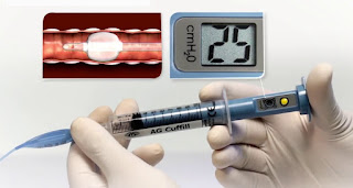
b) AG Cuffill Cuff Inflator with Integrated Manometer: (Figure 2)


AG Cuffill Cuff Inflator with Integrated Manometer
Figure 2: AG Cuffill Cuff Inflator with Integrated Manometer

c) Tru-Cuff ETT Cuff Pressure Syringe: (Figure 3)


Tru-Cuff ETT Cuff Pressure Syringe
Figure 3: Tru-Cuff ETT Cuff Pressure Syringe

d) Vortan Cuff Inflator: (Figure 4)


Vortan Cuff Inflator
Figure 4: Vortan Cuff Inflator

e) AccuCuff Cuff Pressure Indicator: (Figure 5)


AccuCuff Cuff Pressure Indicator
Figure 5: AccuCuff Cuff Pressure Indicator

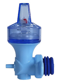
f) PressureEasy Cuff Pressure Controller: (Figure 6)


PressureEasy Cuff Pressure Controller
Figure 6: PressureEasy Cuff Pressure Controller

6-Profile Soft-Seal Cuff (PSSC) ETT:

-The Profile Soft-Seal Cuff (PSSC; Sims Portex, Kent, UK), made of a material impervious to N₂O, velvet soft polyvinyl chloride, a new material with N₂O gas barrier properties produces a thin and highly compliant cuff without increasing N₂O diffusion, thereby reducing the increase of iPcuff and postoperative sore throat.

7-Portex Soft-Seal tube cuff:

-In the Portex Soft-Seal tube cuff (Portex Ltd., Hythe, UK), the plasticizer added to soften the polyvinyl chloride makes the cuff much less permeable to N₂O despite having a thickness of 0.06 mm, which is very similar to the Mallinckrodt Lo-Contour (Athlone, Ireland). The new design prevented increases in the iPcuff and remained stable.

8-Repeated deflation of Air-filled ETT cuff:

-In clinical practice, some anesthesiologists perform a simple method of repeated cuff deflations to inhibit excessive pressure, and eventually, the Pcuff stabilizes. Immediately after TI, the ETT cuff is aspirated as much as possible and then inflated with the smallest volume of air that would produce 12-14 mmHg of Pcuff and seal the airway when the intra-airway plateau pressure is 18 cmH₂O.

-During 67% N₂O anesthesia, gases in the cuff are aspirated every 30 min. in the Trachelon (Terumo, Tokyo, Japan) or 60 min. in the PSSC ETT (Sims Portex, Kent, UK) for 4 h. to decrease the Pcuff to the initial pressure. Pcuff should stabilize when the N₂O concentration is equivalent on both sides of the cuff wall. Therefore, equilibration of the N₂O concentration after repeated deflation for 4 h. is the underlying mechanism of this simple technique. The increased compliance of the PSSC ETT cuff, rather than the N₂O gas barrier, contributes to the requirement for longer intervals between cuff aspirations to avoid excessive pressure compared with what is needed for standard ETTs.

9-N₂O-O₂ gas mixture-filled ETT cuff:

-Immediately after intubation, the cuff is aspirated as much as possible and then inflated with the smallest volume of 40% N₂O and 60% O₂ that would not leak when the intra-airway plateau pressure is 18 cmH₂O. The N₂O gas mixture to fill cuffs is aspirated from the common gas outlet of an anesthetic machine.

-Inflating cuffs with 40% N₂O maintains stable Pcuff without excessive iPcuff or air leaks during anesthesia with 67% N₂O; however, deflationary phenomena of the cuff might occur because iPcuff decreases so quickly after substituting oxygen for N₂O, therefore, it is suggested that the Pcuff be checked frequently to avoid air leaks and aspiration of gastric contents during prolonged emergence from anesthesia or during transportation of patients with TI. 

-The ETT with the N₂O gas-barrier type of cuff might be beneficial because of the longer time required to decrease Pcuff after cessation of N₂O administration and that the highly compliant cuff might be the mechanism of slow changes of iPcuff in the PSSC.

10-Saline-filled ETT cuff:

-When saline is used to fill the cuff, the lack of pressure increase secondary to N₂O diffusion depending on the physical principle that liquids do not expand in volume when highly soluble gases dissolve in them.

11-Lidocaine-filled ETT cuff:

-Lidocaine placed inside the cuff of an ETT can slowly diffuse through its hydrophobic structure. The ETT cuff is prefilled with 7 to 8 ml of L-HCl 2% (140-160 mg) for 90 min. before intubation to enhance the diffusion of lidocaine across the cuff then the cuff is evacuated before intubation. Following intubation, the ETT cuff is inflated with enough lidocaine to prevent retrograde leak at a tidal volume of 10 ml/kg.

12-Alkalinized lidocaine-filled ETT cuff:

-Alkalization of lidocaine can promote the in vitro diffusion across the ETT cuff many tens of times. The cuff of the tracheal tube is initially inflated slowly with saline until no leak is heard under controlled ventilation, after the initial injection of 2 ml of L-HCl 2% (40 mg) into the ETT cuff, a supplementary volume of 2 ml of 8.4% NaHCO₃ is added. It has been shown that small amounts of L-HCl diffused slowly across the ETT cuff; the addition of NaHCO₃ increases the diffusion and dramatically increases the amount of lidocaine released. Plasma lidocaine concentrations increase when lidocaine is alkalinized (Cmax is 62.5 ± 34.0 ng/ml) versus (3.2 ± 1.0 ng/ml) without NaHCO₃. Alkalinization of L-HCl with NaHCO₃ allowed the diffusion of 65% (versus 1% without NaHCO₃) of the neutral base form of lidocaine for 6 hours.

-Use of a small dose of alkalinized lidocaine markedly improves ETT tolerance during a more prolonged time and offers the advantages of minimal stress response to smooth extubation and cough-free emergence. 

Management of Pre-eclampsia

Management of Pre-eclampsia

Pre-eclampsia

➧ The main therapy for pre-eclampsia is to deliver the baby as soon as he/she is most prudent, to enhance maternal and fetal well-being.

1-Magnesium Sulphate (MgSO₂):

➧ Magnesium sulfate has anti-seizure effects as well as being a vasodilator. 

➧ It decreases the pulsatility index in uterine, umbilical, and fetal arteries in women with pre-eclampsia.

➧ It normalizes placental interleukin-6 secretion in a model of pre-eclampsia, which supports the fact that some of its benefits may drive from anti-inflammatory actions. 

➧ The use of MgSO₂ in the management of women with severe pre-eclampsia can reduce the development of eclampsia. However, in women with mild pre-eclampsia, the routine use of MgSO₂ for seizure prophylaxis is not recommended. 

➧ The two most widely used regimens of magnesium sulfate administration are the IV regimen and the IM regimen. 

In the IV regimen: A loading dose of 4 g (usually in 20% solution) is given over 5 min. which is followed by an IV infusion of 1 g/h. for 24 h. after the last seizure.

In the IM regimen: An IV loading dose of 4 g is given over 5 min. followed immediately by 5 g (usually in 50% solution) as a deep IM injection into the upper outer quadrant of each buttock. Maintenance therapy is in the form of a further 5 g IM every 4 h., to be continued for 24 h. after the last fit. If convulsions recur, both regimens advocate a further 2-4 g (depending on the woman’s weight, 2 g if < 70 kg) to be given IV over 5 min.

➧ Parenterally administered magnesium sulfate is cleared almost totally by renal excretion, and magnesium intoxication is avoided by ensuring that urine output is adequate, the patellar or biceps reflex is present, and there is no respiratory depression. 

➧ Eclamptic convulsions are almost always prevented by plasma magnesium levels maintained at 4-7 mEq/L (4.8-8.4 mg/dL or 2-3.5 mmol/L). Patellar reflexes disappear when the plasma magnesium level reaches 10 mEq/L. This sign serves to warn of impending magnesium toxicity. When plasma levels rise above 10 mEq/L, respiratory depression develops, and at 12 mEq/L or more, respiratory paralysis and arrest follow.

2-Antihypertensives:

➧ Due to the risk of hemorrhagic stroke in the presence of systolic hypertension, most guidelines recommend lowering non-severe blood pressure to a systolic level of 140-150 mmHg and a diastolic of 90-100 mmHg. 

➧ Oral safe agents include methyldopa, labetalol, calcium channel blockers (nifedipine or isradipine), and some β-adrenoceptor blockers (metoprolol, pindolol, propranolol) and low-dose diazoxide. 

➧ β-adrenoceptor blockers may cause fetal bradycardia and decrease uteroplacental blood flow.

➧ Atenolol is not recommended due to fetal growth restriction.

➧ Angiotensin-converting enzyme inhibitors (ACEI) and angiotensin II receptor blockers are contraindicated.

➧ Methyldopa is the drug of choice, with well-documented safety after the 1st trimester. It is oral dose is 0.5-3 g/d. in 2 divided doses. 

➧ Labetalol oral dose is 200-1200 mg/d. in 2-3 divided doses.

➧ Nifedipine may inhibit labor and has synergistic interaction with MgSO₄. It is oral dose is 30-120 mg/d. of slow-release preparation.

➧ Medical management of blood pressure should be achieved before obstetric or anesthetic interventions if possible. Control of blood pressure with IV hydralazine, labetalol, or infusions of nitroglycerin or nitroprusside should be commenced with arterial and central venous monitoring in severe cases.

Hydralazine:

➧ It is the drug of choice with long experience in safety and efficacy. Its dose is 5 mg IV or IM, then 5-10 mg every 20-40 min.; or a constant infusion of 0.5-10 mg/h. 

➧ It is also typically employed in more refractory cases.

➧ The use of hydralazine is often accompanied by maternal tachycardia and cautious administration of up to 500 ml crystalloid is recommended before or at the same time as the initial dose of IV hydralazine to reduce the chance of a precipitous fall in blood pressure.

Labetalol:

➧ It produces less tachycardia and arrhythmia than other vasodilators. 

➧ Its dose is 20 mg IV, then 20-80 mg every 20-30 min. up to a maximum of 300 mg; or a constant infusion of 1-2 mg/min.

Glyceryl trinitrate (GTN):

➧ It is the pharmacological agent of choice in women with pre-eclampsia and acute pulmonary edema. 

➧ It is administered as an infusion of 5 µg/min., increasing every 3-5 min. to a maximum dose of 100 µg/min.

Sodium nitroprusside (SNP):

➧ Sodium nitroprusside is rarely used in pregnancy and has known maternal adverse effects of hypotension and paradoxical bradycardia in women with severe pre-eclampsia. 

➧ Fetal cyanide toxicity is a complication of prolonged treatment. SNP should be used with extreme caution in situations of life-threatening hypertension, immediately before delivery in circumstances where clinicians are familiar with its use.

➧ It is administered as an IV infusion at 0.25–5.0 µg/kg/min.

3-Aspirin:

➧ Since inflammation appears to play a significant role in the pathogenesis of pre-eclampsia, benefits from aspirin in the prevention of pre-eclampsia and its vascular complications may derive not just from an anti-inflammatory action but from its effect of restoring the balance between thromboxane and prostacyclin in the vasculature. 

➧ Before using aspirin to prevent pre-eclampsia, consideration must be given to the toxicity in the gastrointestinal tract (GIT) and its effects on renal function.

4-Intravenous fluids:

➧ The use of either crystalloid or colloid solutions has been associated with transient improvements in maternal cardiovascular system parameters. 

➧ Fluid management guided by CVP in severe cases has been demonstrated to improve urine output, maintain mean arterial blood pressure, and decrease diastolic blood pressure. 

➧ If oliguria persists after normalization of CVP (usually 2-3 cm H₂O) or the physiologic state is complicated by pulmonary edema or cardiovascular decompensation, a pulmonary artery catheter (PAC) may be helpful.

➧ A cardiology consultation and an assessment of cardiopulmonary function with a transthoracic echocardiogram may assist with the diagnosis and management.

➧ The use of IV fluids to increase plasma volume or treat oliguria in a woman with normal renal function and stable serum creatinine levels is not recommended. 

➧ Acute pulmonary edema is associated with positive fluid balances of > 5500 mL, which is a frequent cause of admission to intensive care and is a leading cause of death in women with pre-eclampsia.

Complications of Pre-eclampsia

Complications of Pre-eclampsia

Pre-eclampsia

➧ The course of pre-eclampsia can be complicated by mild to severe coagulopathy even in the presence of a normal platelet count.

➧ Severe maternal complications include antepartum hemorrhage due to placental abruption, eclampsia, cerebrovascular accidents, organ failure, and disseminated intravascular coagulation (DIC). 

➧ Deaths are due to intracranial hemorrhage, cerebral infarction, acute pulmonary edema, respiratory failure, and hepatic failure or rupture. 

➧ Eclampsia is the occurrence of seizures in a woman with pre-eclampsia that cannot be attributed to other causes. The seizures are grand mal and may appear before, during, or after labor. Seizures that develop more than 48 h. postpartum may be encountered up to 10 days postpartum. 

➧ Maternal endothelial dysfunction can last for years after the episode of pre-eclampsia. A history of pre-eclampsia is associated with a doubling of the risk for cardiac, cerebrovascular, and peripheral vascular disease compared to women without such a risk factor. Furthermore, such women have an increased risk for renal diseases, such as Focal segmental glomerulosclerosis (FSGS) and microalbuminuria, hypertension, and ischemic heart disease, in later life. 

➧ Pre-eclampsia is the leading cause of fetal growth restriction, intrauterine fetal demise, and preterm birth. 

➧ The children born after pregnancies complicated by pre-eclampsia have also been shown to be at high risk for complications like diabetes mellitus, cardiovascular disease, and hypertension. The pathogenesis of this increased risk has been contributed to fetal malnutrition, epigenetic modification, and postnatal growth acceleration. 

Pathophysiological changes in Pre-eclampsia

Pathophysiological changes in Pre-eclampsia


Pre-eclampsia

1-Cardiovascular changes:

➧ Hypertension in pre-eclampsia is due to marked vasoconstriction because both cardiac output and arterial compliance are reduced.

➧ There is a reversal of the normal circadian rhythm, with the highest blood pressure now at night, and a loss of the normal pregnancy-associated refractoriness to pressor agents; the sensitivity to infused angiotensin II increases weeks before the overt disease.

➧ Increases in insulin resistance and sympathetic nervous system tone also occur and have been implicated in the vasoconstriction characteristic of pre-eclampsia.

➧ The heart may reveal endocardial necrosis similar to that caused by hypoperfusion in hypovolemic shock.

2-Renal changes:

➧ Renal hemodynamics increase markedly in normal gestation, and renal plasma flow (RPF) and GFR decrease in pre-eclampsia (~25%); thus, values may be still above or at those of a non-pregnant state.

➧ The proteinuria of pre-eclampsia is associated with a pathognomonic renal lesion known as glomerular endotheliosis, in which the endothelial cells of the glomerulus swell and endothelial fenestrations are lost.

➧ Podocyturia has been recently associated with pre-eclampsia during clinical disease.

➧ The decrement in RPF is attributable to vasoconstriction, whereas the fall in GFR relates both to the decrement of RPF and the development of glomerular endotheliosis. In rare cases, acute renal failure may develop.

3-Cerebral changes:

➧ There is increased cerebral blood flow in pre-eclamptic women. Cerebral edema and intracerebral parenchymal hemorrhage are common autopsy findings in women who died from eclampsia. However, cerebral edema in eclampsia does not correlate with the severity of hypertension, suggesting that edema is secondary to endothelial dysfunction rather than a direct result of blood pressure elevation.

➧ Findings from head computed tomography (CT) scans and magnetic resonance imaging (MRI) are similar to those seen in hypertensive encephalopathy, with vasogenic cerebral edema and infarctions in the subcortical white matter and adjacent gray matter, predominantly in the parietal and occipital lobes. This syndrome is known as a posterior reversible leuko-encephalopathy syndrome (PRES).

4-Hepatic changes:

➧ Pre-eclampsia also affects the liver. Manifestations include elevated aspartate aminotransferase and lactic dehydrogenase levels, the increments are usually small, except when the HELLP syndrome supervenes.

➧ The gross hepatic changes in pre-eclampsia are petechiae ranging from occasional to confluent areas of infarction, as well as subcapsular hematomas, some having ruptured and caused death.

➧ The characteristic microscopic lesion is periportal, manifesting as hemorrhage into the hepatic cellular columns and at times concurrent infarction.

5-Coagulation changes:

➧ Pre-eclampsia is associated with activation of the coagulation system, with thrombocytopenia (usually mild) as the most commonly detected abnormality.

➧ There is increased platelet activation and size, plus decrements in their lifespan.

➧ The hypercoagulability of a normal pregnancy is accentuated (e.g. reduced antithrombin III, protein S, and protein C) even when platelet counts appear normal.

➧ Occasionally, the coagulopathy can be severe, as in the HELLP syndrome.

6-Eye changes:

➧ The severity of retinal changes depends upon the degree of hypertension. Retinal changes are likely to occur when SBP is above 150 mmHg and DBP is more than 100 mmHg.

➧ Visual disturbances occur including scotoma, diplopia, diminished vision, and photopsia.

➧ The three most common visual complications are hypertensive retinopathy, exudative retinal detachment, and cortical blindness.

➧ Possible explanations for these complications include coexisting or preexisting systemic vascular disease, changes in the hormonal milieu, endothelial damage, abnormal autoregulation, hypoperfusion ischemia, or hyperperfusion edema.

7-Metabolic changes:

➧ These include dyslipidemia with elevated triglycerides, free fatty acids, and low-density lipoprotein (LDL) cholesterol, and reduced high-density lipoprotein (HDL) cholesterol, with an increased prevalence of low dense LDL.

➧ Insulin resistance and uric acid, other components of the metabolic syndrome, are also increased in pre-eclampsia.

8-Utero-Placental changes:

➧ Shallow and abnormal placentation is a hallmark of pre-eclampsia, highlighted by a failure of the normal trophoblastic invasion of the spiral arteries, these vessels fail to remodel and dilate.

➧ This aberration underlies theories that restriction of placental blood flow leads to a relatively hypoxic uteroplacental environment, with subsequent events mediated through hypoxemia-induced genes resulting in the release of factors (e.g. antiangiogenic proteins) that enter the mother’s circulation and initiate the maternal syndrome.

Classification and Pathogenesis of Pre-eclampsia

Classification and Pathogenesis of  Pre-eclampsia


Pre-eclampsia


Classification of Pre-eclampsia:

1. Mild Pre-eclampsia:

Is defined as systolic blood pressure (SBP) of at least 140 mmHg and/or diastolic blood pressure (DBP) of at least 90 mmHg on at least two occasions at least 6 hours apart after the 20th week of gestation in women known to be normotensive before pregnancy and before 20 weeks of gestation plus proteinuria (≥ 300 mg/24 h.). If 24 h. urine collection is not available, then proteinuria is defined as a concentration of at least 30 mg/dL (at least 1+ on dipstick) in at least two random urine samples collected at least 6 hours apart. 

➧ Serum urate levels are often elevated in pre-eclampsia. Hyperuricemia is associated with perinatal complications, and although elevated levels have not predicted adverse maternal outcomes, urate is frequently measured in clinical practice. 

2. Severe Pre-eclampsia:

Is defined as sustained elevations in SBP to at least 160 mmHg and/or in DBP to at least 110 mmHg for at least 6 hours in association with abnormal proteinuria or if there is hypertension in association with severe proteinuria (≥5 g/24 h.). In addition, pre-eclampsia is considered severe in the presence of multiorgan involvement such as pulmonary edema, oliguria (< 500 mL/24 h.), thrombocytopenia (platelet count < 100000 / mm³), abnormal liver enzymes in association with persistent epigastric or right upper quadrant pain, or persistent severe central nervous system (CNS) symptoms (altered mental status, headaches, blurred vision or blindness). 

➧ A severe variant of pre-eclampsia also features hemolysis, elevated liver enzymes, and low platelets (HELLP syndrome). This condition occurs in about 1/1000 pregnancies. Predisposing factors are positive family history, hypertension, diabetes, preexisting renal disease, multiple pregnancies, and poor obstetric history. Pre-eclampsia with hepatic dysfunction, without hemolysis, may occur in severe pre-eclampsia.

Pathogenesis of Pre-eclampsia:

➧ The etiology of preeclampsia remains unknown; however, defective remodeling of spiral arteries during trophoblast invasion is a recognized predisposing factor for pre-eclampsia. Abnormal trophoblast differentiation and incomplete invasion by the placenta of uterine blood vessels leads to the formation of a placenta in which uterine spiral arteries fail to undergo the normal thinning out of muscular walls that permits enhanced perfusion of the placenta. Thus, perfusion of the intervillous space is impaired, leading to placental hypoxia. 

➧ Pre-eclampsia is characterized by placental hypoxia and/or ischemia, excessive oxidative stress, in association with endothelial dysfunction. The release of soluble factors from the ischemic placenta into maternal plasma plays a central role in the ensuing endothelial dysfunction, the most prominent feature of this disease. 

➧ Endothelial dysfunction in pre-eclampsia results from an antiangiogenic state mediated by high circulating levels of soluble Fms-like tyrosine kinase-1 (sFlt-1) and soluble endoglin in concert with low levels of proangiogenic factors like placental growth factor (PlGF), vascular endothelial growth factor (VEGF). The placenta makes sFlt1 in large amounts, but circulating mononuclear cells are an extra source of sFlt1 in pre-eclampsia. 

➧ High-circulating levels of sFlt-1 predate the onset and the severity of pre-eclampsia. Thus sFlt1 acts as a potent inhibitor of VEGF and PIGF by binding these molecules in the circulation and other target tissues, such as the kidneys. Thus excessive sFlt-1 plays a central role in the induction of the pre-eclampsia phenotype as sFLt-1 decreases VEGF binding to its receptor which reduces phosphorylation of endothelial nitric oxide synthase (eNOS) by VEGF leading to reduced eNOS. 

Other Pathophysiologic Mechanisms:

Include immunologic intolerance between fetoplacental and maternal tissues, placental and endothelial dysfunction, immune maladaptation to paternal antigens, exaggerated systemic inflammatory response, maladaptation to the cardiovascular or inflammatory changes of pregnancy, dietary deficiencies, and genetic abnormalities. 

Definition, Differentiation and Risk factors of Pre-eclampsia

Definition, Differentiation, and Risk factors of Pre-eclampsia


Pre-eclampsia

Definition:

➧ Pre-eclampsia is a multi-systemic disease unique to human pregnancy.

➧ The term pre-eclampsia refers to the new onset of hypertension (≥140/90 mmHg) and proteinuria after 20 weeks of gestation in previously normotensive, non-proteinuric women i.e. there is an involvement of one or more organ systems. 

➧ There is a resolution of the disease by three months postpartum i.e. it is specifically a complication of pregnancy. 

➧ Delivery of the baby and removing the placenta is currently the only definitive way of curing the condition. 

➧ The disease is responsible for considerable morbidity and mortality, complicating 5-8% of pregnancies. 

➧ With its systemic vasoconstriction, intravascular volume and protein depletion, and simultaneous retention of extravascular sodium and water, pre-eclampsia is of particular concern to anesthesiologists. 

➧ In addition to individual organ dysfunction, abnormalities in coagulation and edema of the brain, larynx, and lungs may occur.

Atypical Pre-eclampsia:

➧ Atypical cases arise at less than 20 weeks of gestation and over 48 hours after delivery and have signs and symptoms of pre-eclampsia without typical hypertension or proteinuria. 

➧ The condition of pre-eclampsia should be considered, in the absence of proteinuria, when gestational hypertension is present in combination with persistent symptoms or when laboratory tests yield abnormal results.

➧ Pre-eclampsia should be considered in any pregnant woman with a severe headache or new-onset epigastric pain.

➧ Under extremely rare circumstances, pre-eclampsia may develop before 20 weeks of gestation in the setting of a hydatidiform mole, multiple pregnancies, fetal or placental abnormalities, antiphospholipid syndrome, or severe renal disease.

Differentiation of Pre-eclampsia:

➧ Pre-eclampsia is classified within the broad category of hypertensive diseases of pregnancy:

1- Pre-existing or chronic hypertension: is present before and often during pregnancy.

2- Gestational hypertension: is defined as hypertension arising after 20 weeks of gestation, without any other organ system involvement. 

3- Hypertension in pregnancy may be caused by a variety of different pathologies; these include renal disease, pheochromocytoma, drug usage such as cocaine and amphetamines, and cardiovascular diseases such as coarctation, subclavian stenosis, aortic dissection, and vasculitis. 

➧ Other forms of severe hepatic dysfunction in pregnancy need to be differentiated from pre-eclampsia: 

1- Acute fatty liver of pregnancy: is a rare condition of pregnancy, which is not associated with hypertension. 

2- Hemolytic Uremic Syndrome (HUS) ⁄ Thrombotic Thrombocytopenic Purpura (TTP): should be differentiated from HELLP syndrome, as treatment interventions differ significantly. HUS ⁄ TTP is a clinical diagnosis defined by the presence of a pentad of features; microangiopathic hemolytic anemia, thrombocytopenia, neurological symptoms and signs, renal function abnormalities, and fever.

Risk factors:

1- Nulliparity 

2- Multifetal gestations 

3- Previous history of pre-eclampsia 

4- Obesity 

5- Diabetes Mellitus 

6- Vascular and connective tissue disorders like systemic lupus erythematosus and antiphospholipid antibodies 

7- Age >35 years at first pregnancy 

8- Smoking 

9- African-American race 

Accidental Subdural Injection of Local Anesthetics

Accidental Subdural Injection of Local Anesthetics

Predisposing factors:

1-Difficult epidural block. 

2-Previous back surgery. 

3-Recent lumbar puncture (CSF leak through epidural rent leading to distended subdural space). 

4-Rotation of the epidural needle in epidural space through an arc of 180°. 

5-Prolonged epidural catheterization.

Characterized by:

1-Subdural space may not be detected by aspiration test or test dose. 

2-Delayed onset, Short duration. 

3-Extensive block spread (Block is disproportionate to the amount of LA injected). 

4-Segmental, Asymmetric, Patchy distribution. 

5-Cranial spread rather than caudal spread. 

6-High sensory block can involve cranial nerves. 

7-Sparing of sacral roots. 

8-Minimal motor blockade. 

9-Relative lack of sympathetic block. 

N.B. Subdural space has more potential capacity posterior & lateral. There is rarely motor paralysis or severe hypotension due to the sparing of anterior nerve roots that transmit motor & sympathetic fibers.

Detection of subdural placement of epidural catheter:

1-Stimulation test (using nerve stimulator):


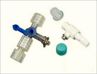
Johns ECG adaptor
Figure 1: Johns ECG adaptor
➧ A nerve stimulator is connected to the epidural catheter via an adapter (Johans ECG adapter, Arrow International, Inc., Reading, USA), (Figure 1). 

➧ The epidural catheter and ECG adapter are primed with sterile normal saline. 

➧ The anode lead of the nerve stimulator is connected to an electrode over the upper or lower extremity as a grounding site. 

➧ The cathode lead of the stimulator is connected to the metal hub of the adapter. 

➧ The nerve stimulator is set at a frequency of 1 Hz with a pulse width of 200 msec. 

➧ Electrical stimulation (1-10 mA) with a segmental motor response (truncal or extremities movement) indicates that the catheter is in the epidural space. 

➧ No motor response indicates that it is not. 

➧ Since it is possible to obtain vigorous and uncomfortable twitches with an excessive current, the current output must be carefully increased from zero and stopped once the motor activity is visible. 

➧ Thus, the stimulator used in the test must be sensitive enough to allow a gradual increase of current output from zero up to at least 10 mA. 

➧ Because a motor response will be elicited at a very low current (< 1 mA) in the case of subarachnoid or subdural placement, the current output must be carefully increased in small increments (0.1 mA) between zero and 2 mA.

2-Contrast Fluoroscopy:

➧ Injection of 3 ml contrast medium via the epidural catheter then radiography of spinal cord is done. 

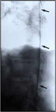
➧ If the epidural catheter is in subdural space, the lateral view will show a contrast medium spreading cephalad over many segments, along the dorsal part of the spinal cord (Figure 2).

Contrast Fluroscopy, Lateral view
Figure 2: Contrast Fluoroscopy, Lateral view