Educational Blog about Anesthesia, Intensive care and Pain management

Ludwig’s Angina

Ludwig’s Angina

Ludwig’s Angina

Definition and Causes:

A rapidly spreading cellulitis of the floor of the mouth that can be produced by any infection. It involves the submandibular, sublingual, and submental spaces.

-Gram-positive cocci (usually streptococci), Staphylococcus aureus, and Staphylococcus epidermidis are now the most common organisms, but sometimes gram-negative rods or anaerobes are responsible. In 50% of cases, more than one organism is isolated.

-It is frequently precipitated by a dental infection involving the second and third lower molars, but trauma may be contributory.

-The condition usually arises from the teeth, but tongue piercing with metal barbells has provided a novel source of infection.

-The frequency is generally decreasing, but there is now a higher incidence in patients with associated systemic diseases. Antibiotics and aggressive surgical treatment have dramatically improved the mortality rate.

-Airway management is controversial, airway compromise develops insidiously, but the actual obstruction is abrupt.

Preoperative Findings:

1. Bilateral submandibular swelling proceeds to brawny induration of the neck. Although the submandibular space is primarily involved in Ludwig’s angina, spread into adjacent fascial spaces may occur.

2. Elevation of the tongue caused by cellulitis of the floor of the mouth.

3. Dysphagia secondary to swelling, and trismus.

4. Gradual onset, or sudden, upper airway obstruction.

5. There is frequently an underlying systemic condition, such as DM, AIDS, or substance abuse.

6. Other complications include; bacteremia, aspiration, retropharyngeal abscess, empyema, mediastinitis, internal jugular vein thrombosis, and pericarditis.

7. Fever, leukocytosis, and increased ESR.

Anesthetic Problems:

1. Trismus, not necessarily relieved by muscle relaxants, may make oral intubation difficult or impossible.

2. Sudden total upper airway obstruction producing hypoxia.

3. Intravenous induction of anesthesia may be hazardous because it can result in apnea and an inability to maintain ventilation on a mask.

4. On a prolonged history of dental sepsis, sepsis may track down through the retropharyngeal space into the posterior mediastinum, requiring awake oral intubation, ventilatory support, and an eventual thoracotomy for a thoracic empyema.

Anesthetic Management:

1. Aggressive early treatment with antibiotics reduces airway problems and the need for surgical intervention.

2. Less severely affected patients may be managed by close observation but only provided that staff is available who can manage acute obstruction. Some argue the case for early tracheostomy.

3. Surgical drainage with or without tooth extraction.

4. Anesthesia for surgery in which there is trismus, but without compromise of the upper airway:

a) Awake fibreoptic nasal intubation but with facilities for emergency tracheostomy available.

b) Inhalation induction and laryngoscopy. If the trismus relaxes and the vocal cords can be seen, a neuromuscular blocker can be given.

c) Tracheostomy may be required in the presence of spreading edema.

5. Airway maintenance in the compromised airway:

-Signs of airway obstruction including; stridor, dyspnea, dysphagia, secretions, and deteriorating oxygen saturations, may indicate the need for rapid, active intervention. In these patients, sedative premedication should be avoided and a drying agent is given.

-If there is significant stridor, a tracheostomy under local anesthesia may be considered.

-Emergency cricothyroidotomy under local anesthesia has also been reported.

Chiari Malformations

 Chiari Malformations

Chiari Malformations


-The Chiari malformation is a term encompassing a range of hindbrain maldevelopments.

-Problems include underdevelopment of the cranial fossa and overcrowding of the normally developed hindbrain, the risks of descent of hindbrain structures below the foramen magnum, and intermittent obstruction to CSF outflow from the fourth ventricle.

-There may be a relatively high CSF pressure in the head and a relatively lower one in the spine.

Types of Chiari Malformations:

There are four types.

Type I:

-Downward herniation of cerebellar tonsils of at least 3–5 mm below the foramen magnum and hindbrain overcrowding results in direct compression of the tissue.

-Clinical symptoms thus relate to CSF disturbances and direct compression of nervous tissue; they include headaches, pseudotumor-like episodes, a syndrome resembling Meniere’s disease, lower cranial nerve signs, and spinal cord disturbances without syringomyelia.

-It is mostly seen in young adults.

Type II (Arnold–Chiari):

-It is the most important and includes; thoracolumbar myelomeningocele, hypoplasia of the posterior fossa, and displacement of the cerebellar vermis, brainstem, and fourth ventricle.

-It presents in infancy or early childhood.

Type III: rare

-Dilated the fourth ventricle with cervical meningomyelocele.

Type IV: rare

-Cerebellar hypoplasia.

Other abnormalities:

1. May be associated with syringomyelia or syringobulbia, other skeletal abnormalities of the skull base and cervical vertebrae, or myelomeningocele. Syringomyelia is the term for an expanding, longitudinal cystic cavity within the spinal cord. In the communicating variety, there is continuity between the syrinx and the CSF in the central canal.

2. May need decompression of foramen magnum or cervical spine.

3. Progressive hydrocephalus may occur which needs shunt surgery.

4. Obstructive sleep apnea.

5. Recurrent pulmonary aspiration.

6. May exhibit abnormal autonomic control.

7. Patients may present with respiratory arrest.

8. Sudden unexpected death may occur.

Anesthetic Problems:

1. Increased ICP may lead to coning.

2. Symptoms may start after a dural puncture.

3. Bolus injections into the epidural space may lead to ICP increases in susceptible individuals.

4. Sleep apnea may occur in Chiari type I.

5. Acute cardiovascular collapse secondary to inadvertent brainstem compression in an infant.

Anesthetic Management:

1. A careful history should be taken in a patient with a ventriculoperitoneal shunt, to check for any signs that it is nonfunctioning. If there is doubt, a referral to a neurosurgeon is indicated. This is particularly important if the notes are absent, there is poor history or lack of follow-up.

2. Try to prevent anything likely to increase ICP, such as coughing, or hypercarbia from heavy sedation or analgesia.

3. Avoid straining in labor. Epidural anesthesia for Cesarean section was performed in a patient with type I Chiari who had a previous difficult tracheal intubation. Small doses of local anesthetic should be titrated gradually, to avoid sudden distension of the epidural space or decreases in arterial pressure.

4. Spinal anesthesia should be avoided.

Budd–Chiari Syndrome

 Budd–Chiari Syndrome

Budd–Chiari Syndrome


-A syndrome caused by obstruction to the hepatic venous outflow and resulting in a clinical picture of hepatomegaly and portal hypertension. It may be secondary to hematological disorders, malignancy, oral contraceptives, heart failure, or constrictive pericarditis.

-The three main sites of obstruction are:

1-Inferior vena cava

2-Large hepatic veins

3-Small intrahepatic venules.

-The condition may be acute or chronic. Acute Budd–Chiari syndrome with hepatocyte necrosis may require urgent portosystemic, decompressive, surgical, or interventional radiological procedures.

-Untreated hepatic venous thrombosis usually results in progressive liver failure and death. Medical treatment is of little help, therefore shunt procedures or liver transplantation will be needed.

Preoperative Findings:

1. Abdominal pain and swelling are the commonest symptoms. Ascites and hepatomegaly will be present in the majority. Vomiting, splenomegaly, jaundice, or bleeding from esophageal varices may also occur. If the vena cava is involved there will be dependent edema.

2. Liver function abnormalities depend on the site and severity of the obstruction. A liver biopsy may show outflow obstruction, hepatic necrosis, and fibrosis.

3. Etiological factors include polycythemia, paroxysmal nocturnal hemoglobinuria, antiphospholipid syndrome, protein C deficiency, factor V Leiden, myeloproliferative disorders, and mechanical factors, such as webs and tumors. Hepatic venous thrombosis has been reported in association with ulcerative colitis, but in the patient described, iron deficiency had concealed an underlying polycythemia vera.

4. Treatment is aimed at the preservation of liver function and includes thrombolysis, angioplasty, stent placement, portacaval shunt, mesoatrial shunt, and liver transplantation.

Anesthetic Problems:

1. Hepatic function may be compromised.

2. Surgery may be required for portacaval or mesoatrial shunt or liver transplantation. This has been described in a patient with paroxysmal nocturnal hemoglobinuria.

3. Patients may present during pregnancy when the condition must be distinguished from HELLP syndrome and acute fatty necrosis of the liver. Antiphospholipid syndrome and preeclampsia in a primipara presented with Budd–Chiari syndrome from thrombosis of the right hepatic veins.

Thrombotic thrombocytopenic purpura resulted in postpartum hepatic venous thrombosis. Paroxysmal nocturnal hemoglobinuria has been associated with hemolytic crises and Budd–Chiari syndrome during pregnancy. Maternal deaths have been reported.
Anesthetic Management:

1. Hematological examination is important because, in the absence of mechanical causes for hepatic vein thrombosis, there is a high incidence of underlying hematological abnormalities; these include myeloproliferative disorders, and paroxysmal nocturnal hemoglobinuria, systemic lupus erythematosus, and antithrombin III deficiency.

2. Assessment of liver function, including coagulation.

3. Low-dose heparin infusion from the first postoperative day has been recommended, with subsequent low-dose aspirin and long-term anticoagulation to reduce the risks of graft thrombosis.

4. Liver transplantation will cure the inherited thrombophilias, for example, factor V Leiden, protein C and S, etc.

5. In a patient with paroxysmal nocturnal hemoglobinuria who developed Budd–Chiari syndrome, the resolution was achieved following bone marrow transplantation.

6. Pregnant patients with paroxysmal nocturnal hemoglobinuria should be carefully monitored for the onset of Budd–Chiari syndrome.

Tachycardia during Anesthesia

Tachycardia during Anesthesia


Tachycardia


Definition:

-Pulse rate greater than 100 beats/min. in adults

Causes and Management:

1-Light anesthesia, Pain:

-Hypertension, sweating, lacrimation, reactive pupils, movement 

Treatment: Deepening the anesthesia, Analgesia 

2-Drug induced:

-Anticholinergic drugs, Catecholamines, Oxytocin 

3-Hypovolemia:

-Actual (due to fluid loss): 

Treatment: Fluid replacement 

-Effective (due to vasodilatation): 

Treatment: Vasopressors (α-agonists, Ephedrine) 

4-Hypercarbia:

Treatment: Check soda lime, Increase minute ventilation, and Exclude malignant hyperthermia 

5-Hypoxia:

The initial response is tachycardia 

Treatment: Of the cause

6-Cardiac dysrhythmia:

-SVT: 

Treatment: Of the cause, Carotid sinus massage, Adenosine, Verapamil, Amiodarone, Digoxin 

-VT: 

Treatment: Of the cause, Lidocaine, Amiodarone, Synchronized DC (if sustained or hemodynamically unstable) 

7-Endocrine disease:

-Pheochromocytoma, Thyrotoxic crisis 

Treatment: α-blockers, β-blockers 

8-Sepsis:

9-Malignant hyperthermia:

The first sign is tachycardia 

Treatment: According to management guidelines

Bradycardia during Anesthesia

Bradycardia during Anesthesia

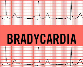
Bradycardia

Definition:

-Pulse rate less than 60 beats/min. in adults

-Pulse rate less than 80 beats/min. in infants

-Pulse rate less than 100 beats/min. in neonates

Causes and Management:

1-Hypoxia:

-Late response is bradycardia

Treatment:

-Of the cause, Oxygenation, Anticholinergics

2-Drug induced:

-High-concentration volatile anesthetics, Opioids, Succinylcholine, Anticholinesterases (Neostigmine), Low dose atropine (Benzold-Jarisch reflex, paradoxical bradycardia), β-blockers, Digoxin

Treatment:

-Decrease concentration of volatile anesthesia, Anticholinergics

3-Vagal stimulation:

- Airway instrumentation, Visceral traction, Extraocular muscle traction, Anal dilatation, Cervical dilatation

Treatment:

-Stop traction or dilatation, Anticholinergics

4-Spinal anesthesia:

-High spinal anesthesia affecting T1-T4 (Cardiac accelerator fibers)

Treatment:

-Support circulation, Anticholinergics, Ephedrine (if associated with hypotension)

5-Ischemic heart disease:

-Ischemic changes affecting the conducting system

Treatment:

-Anticholinergics if indicated

6-Endocrine disease:

-Hypothyroidism

Treatment:

-Anticholinergics

7-Metabolic:

-Hyperkalemia

Treatment:

-Correction of potassium level, Anticholinergics

8-Neurological:

-Cushing’s reflex due to increased ICP

Treatment:

-Of the cause, Anticholinergics if indicated

9-Cardiovascular fitness:

-Trained athlete (High resting vagal tone, Large stroke volume)

N.B.: Anticholinergic drugs (Atropine, Glycopyrrolate, Hyoscine)


Esophageal Achalasia

Esophageal Achalasia

Esophageal Achalasia

Definition:

A chronic, progressive motor disorder of the esophagus associated with degenerative changes in the myenteric ganglia and vagal nuclei.

Components:

There are three components:

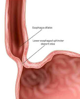
1-Failure of the lower esophageal sphincter to relax, with an increased resting sphincter pressure, which together results in a functional obstruction

2-Absence of sequential peristalsis in response to a bolus of food

3-Dilated, contorted esophagus

Pathophysiology and Management:

-Degeneration of the myenteric plexus and decreased nitric oxide synthesis may be the problem.

-Overspill may produce bronchopulmonary complications, and 5–10% of patients ultimately develop carcinoma of the esophagus.

-Nitrates and calcium channel blockers given before meals sometimes produce symptomatic improvement, but the mainstays of treatment are esophageal dilatation and surgical myotomy.

-Open surgery has been mostly replaced by laparoscopic myotomy and fundoplication.

-For elderly patients, an endoscopic injection of botulinum toxin can give relief for several months without the risk of surgery.

Preoperative Findings:

1. Symptoms include; dysphagia, retrosternal pain, regurgitation, and weight loss. In young people, the condition may be misdiagnosed as anorexia nervosa or asthma.

2. Respiratory complications, which may be attributed to asthma or chronic bronchitis, are secondary to the overspill of undigested material.

3. Nocturnal coughing occurs in 30%, and bronchopulmonary complications in 10% of patients.

4. The aspiration of larger volumes may result in lobar collapse, bronchiectasis, or lung abscess.

5. Rarely, it may present with a cervical mass and acute upper respiratory tract obstruction, necessitating urgent intervention.

6. There is an increased risk of esophageal carcinoma.

7. Diagnosis can be made on barium swallow, manometric studies, and endoscopy. Occasionally, acute dilatation may be seen on CXR, in which case, abnormal flow–volume curves will indicate variable intrathoracic tracheal obstruction.

Anesthetic Problems:

1. A predisposition to regurgitation and pulmonary aspiration in the perioperative period.

2. Passage of the tracheal tube past the dilated esophagus can be achieved with difficulty.

3. During recovery from anesthesia, neck swelling, and venous engorgement can be precipitated by coughing or straining. Acute thoracic inlet obstruction with stridor, deep cyanosis of the face, and hypotension can occur.

4. Upper airway obstruction or respiratory failure, particularly in the elderly. Rarely, an acute dilatation of the esophagus results in total airway obstruction.

5. Acute respiratory failure can occur after surgery.

6. The opening pressure of the cricopharyngeus muscle from above is much lower than that from below, therefore progressive dilatation of the upper esophagus may occur, particularly in association with mask ventilation or IPPV.

7. An increased intrathoracic pressure produced by a Valsalva maneuver forces air from the thoracic into the cervical esophagus. Occasionally, death can occur.

8. If acute airway obstruction is present, sudden decompression of the esophagus may cause the pharynx to flood with food and fluid, resulting in aspiration.

Anesthetic Management:

1. If anesthesia is required, precautions must be taken to reduce the risk of aspiration of gastric contents. The dilated esophagus must be emptied and decompressed. This needs a period of prolonged starvation, possibly with washouts of the esophagus.

2. A rapid sequence induction should be undertaken with awake tracheal extubation, the patient should be nursed in the lateral position during recovery.

3. Sublingual nifedipine 10–20 mg has been shown to reduce the basal sphincter pressure after 10 min and the effect lasts for up to 40 min.

4. Management of acute upper airway obstruction secondary to tracheal compression has been reported using the following methods:

a) Sublingual glyceryl nitrate.

b) Passage of a naso-esophageal tube.

c) Transcutaneous needle puncture.

d) Tracheal intubation.

e) Rigid esophagoscopy.

f ) Emergency tracheostomy.

g) Cricopharyngeus myotomy.

5. Treatment can be either surgical or medical. For the elderly and less fit patients, pneumatic dilatation, or endoscopic injection of botulinum toxin, may be appropriate. Heller myotomy and partial fundoplication can be performed either as an open or a laparoscopic procedure.

Fat Embolism Syndrome (FES)

Fat Embolism Syndrome (FES)

➧ Fat embolism can be difficult to diagnose. It most often follows a closed fracture of a long bone but there are many other causes.

Epidemiology:

➧ Incidence of this complication ranges from 0.5 to 11% in different studies. It varies considerably according to the cause. Patients with fractures involving the middle and proximal parts of the femoral shaft are more likely to experience fat embolism. Age also seems to be a factor with young men at the highest risk.

Etiology:

1-Fractures: Closed fractures produce more emboli than open fractures. Long bones, pelvis, and ribs cause more emboli. Sternum and clavicle cause less. Multiple fractures produce more emboli.

2- Orthopedic Procedures: Intramedullary nailing of long bones, Fixation of cemented hip prosthesis.

3-Massive soft tissue injury

4-Severe burns

5-Liposuction

6-Bone marrow biopsy

7-Bone marrow harvesting and transplant

8-Cardio-pulmonary bypass

9-Non-traumatic settings occasionally lead to fat embolism. These include conditions associated with: 

-Fatty liver

-Acute pancreatitis

-DM

-Osteomyelitis

-Bone tumor Lysis

-Pathological fractures

-Sickle cell crisis (causing bone infarcts)

-Decompression sickness

-Prolonged corticosteroids therapy

-Cyclosporine A solvent

-Parenteral lipid infusion

Sevitt’s classification:

1-Subclinical (Incomplete) form: Fat emboli present in blood and lungs, no symptoms/signs

2-Non-fulminant (Classic) form: Pulmonary dysfunction, cerebral dysfunction, petechiae

3-Fulminant form: Rare, rapid onset (within hours) of acute cor-pulmonale, respiratory failure, coma, and death.

Principal clinical features (triad) of FES:

1-Respiratory failure (dyspnea and hypoxia)

2-Cerebral dysfunction (confusion)

3-Petechiae

➧ Clinical features usually present between 24-72 h. after trauma (lucid interval). This is especially after fractures, when fat droplets act as emboli, becoming impacted in the pulmonary microvasculature and other microvascular beds, especially in the brain.

➧ Embolism begins rather slowly and attains a maximum in about 48 h.

➧ The initial symptoms are probably caused by mechanical occlusion of multiple blood vessels with fat globules that are too large to pass through the capillaries.

➧ The vascular occlusion in fat embolism is often temporary or incomplete as fat globules do not completely obstruct capillary blood flow because of their fluidity and deformability.

➧ The late presentation is thought to be a result of hydrolysis of the fat to more irritating free fatty acids which then migrate to other organs via the systemic circulation.

➧ It has also been suggested that paradoxical embolism occurs from shunting.

Clinical Presentation:

➧ There is usually a latent period of 24-72 h. between injury and onset.

➧ Symptoms: There is vague pain in the chest and shortness of breath.

➧ Signs: The onset is sudden, with:

-Restlessness

-Fever occurs, often at more than 38.3° C with a disproportionate tachycardia.

-Drowsiness with oliguria is almost pathognomonic.

➧ CNS signs: including a change in the level of consciousness, are common. They are usually non-specific and have the features of diffuse encephalopathy with acute confusion, stupor, coma, rigidity, or convulsions. Cerebral edema contributes to neurological deterioration.

Diagnostic Criteria:

A) Gurd's and Wilson’s Criteria:

-Diagnosis requires at least one sign from the major criteria and at least four signs from the minor criteria. (Table 1)


Gurd's and Wilson’s Criteria
Table 1: Gurd's and Wilson’s Criteria

B) Schönfeld’s Criteria:

-Diagnosis requires a score of more than 5. (Table 2)


Schönfeld’s Criteria
Table 2: Schönfeld’s Criteria

C) The clinical diagnosis is assured if all three of the following criteria are present within 72 h. after traumatic fracture:

1-Unexplained dyspnea, tachypnea, arterial hypoxia with cyanosis, and diffuse alveolar infiltrates on chest X-ray.

2-Unexplained signs of cerebral dysfunction, such as confusion, delirium, or coma.

3-Petechiae over the upper half of the body, conjunctiva, oral mucosa, and retina.

Differential Diagnosis:

➧ Dyspnea, hypoxia, and abnormal CXR: can occur with thrombo-embolism and pneumonia.

➧ Cerebral dysfunction: can occur with hypoxia or meningitis but the rash of meningococcal septicemia is all over and spreads rapidly. It can also occur as a late feature of head injury.

➧ Thrombotic Thrombocytopenic Purpura (TTP).

Investigations:

CBC: 

-Hematocrit: is decreased, occurs within 24-48 h., and is due to intra-alveolar hemorrhage. 

-Platelets: are decreased.

Serum Lipase: is increased, but this is not pathognomonic as it occurs in any bone trauma.

Serum Calcium: is decreased.

Cytological Examination: urine, blood, and sputum may detect fat globules that are either free or in macrophages. This test has low sensitivity and a negative result does not exclude fat embolism. 

Arterial Blood Gases: will show hypoxia, PaO₂ usually less than 60 mmHg, and hypocapnia. Continuous pulse oximeter monitoring may enable hypoxia from fat embolism to be detected in at-risk patients before it is clinically apparent.

ECG: Ischemia, Rt. Ventricular strain, Rt. Axis deviation, RBBB, Arrhythmia.

Chest X-ray: may show evenly distributed, fleck-like pulmonary shadows (Snow Storm appearance), increased pulmonary markings, and dilatation of the right side of the heart.

CT Head: (to rule out intracranial pathology).

CT Chest: (to rule out chest pathology).

Management:

A) Supportive

1-Ensuring good arterial oxygenation: High flow rate of oxygen is given to maintain the arterial oxygen tension in the normal range.

2-Restriction of fluid intake and Diuretics: to minimize fluid accumulation in the lungs so long as circulation is maintained.

3-Maintenance of intravascular volume: is important because shock can exacerbate the lung injury caused by FES. Albumin has been recommended for volume resuscitation in addition to the balanced electrolyte solution, because it not only restores blood volume but also binds fatty acids, and may decrease the extent of lung injury.

4-Mechanical ventilation (APRV) and Positive End-Expiratory Pressure (PEEP): may be required to maintain arterial oxygenation.

B) Pharmacological

1-Corticosteroids: Methylprednisolone (membrane stabilizer, decreases endothelial damage caused by free fatty acids).

2-Low dose heparin: 2500 u / 6 h. (reduce the degree of pulmonary comprise and intravascular coagulation despite the risk of hemorrhage and intravascular lipolysis).

3-Dextran-40: (decreases intravascular thrombosis when ESR is elevated).

4-Ethanol: (decreases lipolysis)

5-Dextrose: (decreases free fatty acid mobilization)

6-DVT prophylaxis

7-Nutrition

C) Surgical:

1-Prompt surgical stabilization of long bone fractures within 24 h. reduces the risk of the syndrome.

2-Use of vacuum or venting during reaming of long bones.

3-Prophylactic IVC filter in at-risk patients.

Prognosis:

➧ The mortality rate from FES is 5-15%. Even severe respiratory failure associated with fat embolism seldom leads to death.

➧ The prognosis is worse in older patients and those with more severe injury but is not affected by gender.

➧ Criteria of bad prognosis: Sevitt’s class, Serum lipase, Lipuria, ARDS.

Prevention:

➧ Early immobilization of fractures seems to be the most effective way of reducing the incidence of this condition.

Porphyric Crisis

Porphyric Crisis (Acute Neurovisceral Crisis) 

Background: 

-The porphyrias are caused by enzyme deficiencies in the heme production pathway. Such deficiencies may be due to inborn errors of metabolism or exposure to environmental toxins or infectious agents.

-The disease was named porphyria due to the red discoloration of urine in affected patients, (Figure 1).

-Crises are four to five times more common in women and usually occur in their early 30s.

Porphyria red urine
Figure 1: Porphyria Red Urine

Triggering factors: 

Enzyme-inducing drugs: 

-Barbiturates (Thiopental, Methohexital), Etomidate, Enflurane, Alcuronium, Mepivacaine, Pentazocine, Nifedipine, Verapamil, Diltiazem, Phenytoin, Hydralazine, Phenoxybenzamine, Aminophylline 

Physiological: 

-Menstruation, Fasting, Dehydration, Stress, Infection, Anemia, Endogenous hormones 

Habits: 

-Smoking, Alcohol 

Clinical Picture: 

CNS: 

-Autonomic neuropathy (Fever, Pain, Constipation, Gastroparesis, Postural hypotension) 

-Peripheral neuropathy (Skeletal muscle weakness, Quadriparesis, Bulbar palsy, Respiratory failure) 

-Cranial nerve palsy 

-Seizures 

-Psychiatric features (Mood disturbance, Confusion, Psychosis) 

CVS: 

-Tachycardia, Hypertension 

GIT: 

Acute abdominal pain, Vomiting, Diarrhea, Dehydration, Electrolyte disturbance (↓ [Na⁺, K⁺, Mg⁺²]) 

Management: 

Remove triggering factors (above) 

Specific R: 

-Hematin, Heme arginate / Heme albumin, Somatostatin, Plasmapheresis 

Symptomatic R: 

-Pain: Substantial doses of Opioids 

-Nausea and Vomiting: Prochlorperazine, Ondansetron 

-Anxiety: Lorazepam, Midazolam in low doses 

-Insomnia: Zopiclone 

-Delirium: Haloperidol 

-Tachycardia & Hypertension: β-adrenergic blocking agents, Glyceryl trinitrate 

-Seizures: Benzodiazepines, Levetiracetam, Clonazepam, Gabapentin, Vigabatrin, Magnesium sulphate 

-Sedation: Propofol, Alfentanil infusions. The clinical safety of prolonged midazolam infusion is unknown. 

-Thromboembolic prophylaxis: LMW heparins 

-Stress ulcer prevention: IV Omeprazole, Ranitidine 

-Correction of electrolytes

Mechanical Ventilation for Respiratory failure

Treacher Collins Syndrome

Treacher Collins Syndrome:


Treacher Collins Syndrome
-A craniofacial defect associated with developmental anomalies of the first arch. 

-Abnormalities vary from minimal, to complete syndrome. 

-The syndrome is named after Edward Treacher Collins, an English surgeon and ophthalmologist, who described its essential traits in 1900. 

-Patients may require anesthesia for maneuvers to improve upper airway obstruction temporarily, or for correction of some of the congenital defects. 

-Airway obstruction and a requirement for multiple operations increase the need for tracheostomy. 

Etiology:

-It is due to mutations in TCOF1, POLR1C, or POLR1D genes. TCOF1 gene mutations are the most common cause of the disorder, accounting for 81 to 93% of all cases. POLR1C and POLR1D gene mutations cause an additional 2% of cases. In individuals without an identified mutation in one of these genes, the genetic cause of the condition is unknown. 

Preoperative Abnormalities:

1. Features may include mandibular and malar hypoplasia, antimongoloid palpebral fissure, macrostomia, irregular maloccluded teeth, microphthalmia, lower lid defects, cleft palate, high arched palate, macroglossia, and auricular deformities. 

2. Associated abnormalities include mental retardation, deafness, dwarfism, cardiac defects, choanal atresia, and skeletal deformities. 

3. Chronic upper respiratory tract obstruction and obstructive sleep apnea, which can lead to growth retardation and/or cor-pulmonale. 

Anesthetic Problems:

1. Upper airway obstruction: in neonates, this may require urgent temporary maneuvers, such as stitching the tongue to the lower lip 

2. Excess secretions: may impede induction of anesthesia 

3. Inhalational induction: may be difficult 

4. Difficult tracheal intubation 

5. Obstructive sleep apnea may occur postoperatively 

6. Pulmonary edema 

Intraoperative Management:

Recommendations:

1-Monitoring by pulse oximetry, to detect airway obstruction, is crucial. 

2-Avoid respiratory depressant drugs, both for premedication and postoperatively 

3-Use drying agents 

4-Never give muscle relaxant until the airway has been secured. 

Airway Management:

-Several methods have been proposed to overcome the problem of difficult intubation, some in the awake patient and some under general anesthesia: 

a) In Awake Patients: 

1-Awake intubation or awake direct laryngoscopy to visualize the vocal cords. 

2-Direct laryngoscopy, with the patient in the sitting position, and using a 5-G feeding tube taped to the side of the laryngoscope to give oxygen. 

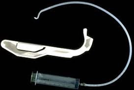
3-The Augustine guide (Figure 1): can be used for nasotracheal intubation in an awake, sedated patient.

Augustine guide
Figure 1: Augustine guide

4-Fibreoptic bronchoscope or Tracheostomy under local anesthesia: 

In small infants, the ‘tube over bronchoscope’ technique is not always possible because of the small size of the tube, therefore a Seldinger-type approach may be necessary: 

-After the administration of atropine, ketamine IM, and topical lidocaine, a fibreoptic bronchoscope (OD 3.6mm, L 60 cm, and suction channel 1.2 mm) was passed through one nostril. 

-The tongue was held forward with Magill forceps, until the vocal cords were seen, but not entered, because of the risk of total obstruction. 

-Under direct vision, a Teflon-coated guidewire with a flexible tip was passed via the suction channel into the trachea. 

-The bronchoscope was carefully removed leaving the wire in place, and an ID 3mm nasotracheal tube was then passed over it into the trachea. 

Pediatric bronchoscopes of 2.5 mm diameter are now available, but their very fineness makes them less easy to handle than the 4-mm bronchoscopes). 

b) In Anesthetized Patients: 

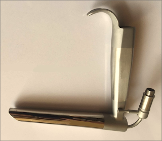
1-Jackson anterior commissure laryngoscope (Figure 2): 

-The head is elevated above the shoulders, with flexion of the lower cervical vertebrae and extension at the atlanto-occipital joint. The laryngoscope is introduced into the right side of the mouth. Only the tip is directed towards the midline, the proximal end remaining laterally so that a further 30 degrees of anterior angulation can be obtained. 

-The narrow, closed blade prevents the tongue from falling in and obscuring the view of the larynx. When visualized, the epiglottis is elevated, and the larynx is entered. Intubation is then achieved by passing a lubricated tube, without its adaptor, down the laryngoscope. It is held in place with alligator forceps whilst the laryngoscope is withdrawn.

Jackson anterior commissure laryngoscope
Figure 2: Jackson anterior commissure laryngoscope

2-The Bullard intubating laryngoscope (Figure 3):

-It can be used to achieve nasotracheal intubation.

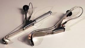
Bullard intubating laryngoscope
Figure 3: Bullard intubating laryngoscope

3-Tactile nasal intubation: 

-Inhalational induction with sevoflurane, and the tongue is pulled downwards and forwards. The tube is initially used as a nasal airway, whilst the index and middle finger are used to palpate the epiglottis, through which the tube is then passed. 

4-Laryngeal mask airway: 

-Inserted under propofol anesthesia and can be used as a conduit for the passage of a fibreoptic bronchoscope.

5-The use of an assistant to pull out the tongue with Magill forceps, and at the same time to apply cricoid pressure, to assist laryngoscopy. 

Postoperative Management:

-The tracheal tube should remain in place until the patient is fully awake. 

-Patients should be nursed in a high-dependency area postoperatively. The combination of sleep apnea and drugs with CNS-depressant effects may make them particularly susceptible to respiratory arrest.

Read more ☛ Pierre Robin Syndrome