Educational Blog about Anesthesia, Intensive care and Pain management

Ebstein’s Anomaly

Ebstein’s Anomaly


-A rare congenital cardiac abnormality.

-The septal and posterior cusps of the tricuspid valve are displaced downwards and are elongated, such that a varying amount of the right ventricle effectively forms part of the atrium. Its wall is thin and it contracts poorly. The remaining functional part of the right ventricle is therefore small.

-The foramen ovale is patent, or defective, in 80% of cases.

-The degree of abnormality of right ventricular function, and the size of the ASD, are probably the main determinants of the severity of the condition, which varies considerably.

-The right ventricular systolic pressure is low, and the RVEDP is elevated. Tricuspid incompetence can occur.

-There may be a right to left shunt, with cyanosis, on effort, and pulmonary hypertension, and right heart failure may supervene.

-The natural history of the disease is very variable. Fifty percent of cases present in infancy with cyanosis, and 42% die in the first 6 weeks of life.

-In those who survive to adulthood, symptoms may be precipitated by the onset of arrhythmias, or by pregnancy. A few patients remain asymptomatic, even as adults, although once symptoms develop, the disability can increase rapidly.

-A cardiothoracic ratio of ≥ 0.65 is a better predictor of sudden death than the symptomatic state, and those who developed atrial fibrillation died within 5 years. It has therefore been suggested that tricuspid surgery should be undertaken before the cardiothoracic ratio reaches 0.65.

Preoperative Abnormalities:

1. There may be a right to left shunt, with dyspnea and cyanosis at rest, or on moderate exertion. Alternatively, the patient may be asymptomatic.

2. Episodes of tachyarrhythmias occur in 25% of patients. Some provoke syncopal attacks.

3. The ECG may show varying abnormalities, including large peaked P waves, a long P–R interval, Wolff–Parkinson–White syndrome, RBBB, and right heart strain. Paroxysmal supraventricular tachycardia occurs in 15%, usually because of the presence of WPW syndrome.

4. Chest X-ray may show cardiomegaly, with a prominent right heart border, and poorly perfused lung fields.

5. Paradoxical systemic embolism and bacterial endocarditis may occur.

6. Many other lesions of the tricuspid valve or right ventricle may mimic Ebstein’s anomaly, therefore the discriminating clinical and echocardiographic features for correct diagnosis have been enumerated.

Anesthetic Problems:

These will depend upon the anatomical abnormality, the degree of right to left shunt, and the presence or absence of right heart failure.

1. Induction time is prolonged, because of the pooling of drugs in the large atrial chamber.

2. Intracardiac catheter insertion may be hazardous because it can provoke serious cardiac arrhythmias.

3. Air entering peripheral venous lines or any open veins at subatmospheric pressure may cause paradoxical air emboli.

4. Tachycardia is poorly tolerated because of impaired filling of the functionally small right ventricle.

5. Hypotension may increase the right to left shunt if present.

6. Hypoxia causes pulmonary vasoconstriction, which also increases a right to left shunt.

7. There is a risk of bacterial endocarditis, especially if a CVP line is in place.

8. Deterioration may occur in pregnancy because of a decrease in right ventricular function, and an increase in blood volume and cardiac output, or with the onset of arrhythmias.

Management:

1. The severity of the lesion must be assessed. In the presence of maternal cyanosis or arrhythmias during pregnancy, there should be close monitoring of both mother and fetus. Deterioration may occur, despite previous successful pregnancies.

2. Treatment of heart failure and arrhythmias.

3. Antibiotic prophylaxis against bacterial endocarditis.

4. If a CVP is used for monitoring, its tip should be kept within the superior vena cava. The use of intracardiac catheters should probably be avoided.

5. Techniques should aim to minimize tachycardia and hypotension.

6. Oxygen therapy increases pulmonary vasodilatation. Long-term maternal therapy is required during pregnancy from 14 weeks, to treat fetal hypoxia that is demonstrated by umbilical venous blood gases.

7. Several anesthetic techniques have been described. A two-catheter epidural technique can be used for vaginal delivery to minimize hypotension. Bupivacaine doses must be fractionated and saline rather than air used to site the epidural, to avoid paradoxical air emboli. Cesarean section under general anesthesia, preceded by fentanyl, and a neurolept analgesic technique for hysterectomy, have been described.

Ovarian Hyperstimulation Syndrome (OHSS)

Ovarian Hyperstimulation Syndrome (OHSS)

- It is a complication of some forms of fertility medication. Most cases are mild, but small proportions are severe.


Causative Medications:

- Associated with the injection of a hormone called human chorionic gonadotropin (hCG) which is used for inducing final oocyte maturation and/or triggering oocyte release.
- The risk is further increased by multiple doses of hCG after ovulation and if the procedure results in pregnancy.
- Using a gonadotropin-releasing hormone (GnRH) agonist instead of hCG for inducing final oocyte maturation and/or release results in an elimination of the risk of OHSS, but a slight decrease of the delivery rate of approximately 6%.

Epidemiology:

- Sporadic OHSS is very rare and may have a genetic component.
- Clomifene citrate therapy can occasionally lead to OHSS, but the vast majority of cases develop after the use of gonadotropin therapy (with the administration of FSH), (Menopur, Pergonal, Repronex) and administration of hCG to induce final oocyte maturation and/or trigger oocyte release, often in conjunction with in vitro fertilization (IVF).
- The frequency varies and depends on patient factors, management, and methods of surveillance. About 5% of treated patients may encounter moderate to severe OHSS.
- Mortality is low, but several fatal cases have been reported.

Risk Factors:

- Risk factors include young age, the development of many ovarian follicles under stimulation, extremely elevated serum estradiol concentrations, the use of hCG for final oocyte maturation and/or release, the continued use of hCG for luteal support, and the occurrence of a pregnancy (resulting in hCG production).

Pathophysiology:

- OHSS has been characterized by the presence of multiple luteinized cysts within the ovaries leading to ovarian enlargement and secondary complications.
- The central feature of clinically significant OHSS is the development of vascular hyperpermeability and the resulting shift of fluids into the third space.
- As hCG causes the ovary to undergo extensive luteinization, large amounts of estrogens, progesterone, and local cytokines are released. It is almost certain that vascular endothelial growth factor (VEGF) is a key substance that induces vascular hyperpermeability, making local capillaries "leaky", leading to a shift of fluids from the intravascular system to the abdominal and pleural cavity.
- Supraphysiologic production of VEGF from many follicles under the prolonged effect of hCG appears to be the specific key process underlying OHSS. Thus, while the patient accumulates fluid in the third space, primarily in the form of ascites, she actually becomes hypovolemic and is at risk for respiratory, circulatory, and renal problems. Patients who are pregnant sustain the ovarian luteinization process through the production of hCG.
- Avoiding OHSS typically requires interrupting the pathological sequence, such as avoiding the use of hCG. One alternative is to use a GnRH agonist instead of hCG.

Classification:

Mild OHSS:

- The ovaries are enlarged (5–12 cm), slight weight gain, mild abdominal distension, abdominal pain, nausea, diarrhea, and there may be an accumulation of ascites.

Moderate OHSS:

- Excessive weight gain (> 2 pounds/day or > 1 kg/day), increased abdominal girth, vomiting, darker urine and oliguria, excessive thirst, and dry skin and/or hair (in addition to mild symptoms).

Severe OHSS:

- Marked abdominal distention, lower abdominal pains, darker urine or anuria, pleural effusion, respiratory distress, calf and chest pains. There may be hemoconcentration, and thrombosis (in addition to mild and moderate symptoms).
- Symptoms generally resolve in 1 to 2 weeks but will be more severe and persist longer if pregnancy occurs. This is due to hCG from the pregnancy acting on the corpus luteum in the ovaries in sustaining the pregnancy before the placenta has fully developed. Typically, even in severe OHSS with a developing pregnancy, the duration does not exceed the first trimester.
- Early OHSS develops before pregnancy testing and late OHSS is seen in early pregnancy.

Criteria for Severe OHSS:

- Enlarged ovary, ascites.
- Hematocrit > 45%, WBC > 15,000/microliter.

- Oliguria, creatinine 1.0-1.5 mg/dL, creatinine clearance > 50 ml/min.
- Liver dysfunction, anasarca (extreme generalized edema).

Critical OHSS:

- Enlarged ovary, tense ascites with hydrothorax and pericardial effusion.
- Hematocrit > 55%, WBC > 25,000/microliter.
- Oligoanuria, creatinine > 1.6 mg/dL, creatinine clearance < 50 mL/min.
- Renal failure, thromboembolic phenomena (DVT, PE), ARDS.

Complications:

- Ovarian torsion, ovarian rupture, thrombophlebitis, and renal insufficiency.

Prevention:

- Physicians can reduce the risk of OHSS by monitoring FSH therapy, and by withholding hCG medication.
- Regarding dopamine agonists as prophylaxis, a systematic review and meta-analysis concluded that prophylactic treatment with Cabergoline (Dostinex) reduces the incidence, but not the severity of OHSS, without compromising pregnancy outcomes.
- OHSS may also be prevented by coasting, which is ovarian hyperstimulation in IVF without hCG administration for final maturation of follicles.

Treatment:

Mild OHSS:

- Can be treated conservatively with monitoring of abdominal girth, weight, and discomfort on an outpatient basis until either conception or menstruation occurs. Conception can cause mild OHSS to worsen in severity.

Moderate OHSS:

- Is treated with bed rest, fluids (IV hydration), and close monitoring of labs such as electrolytes (hyponatremia, hyperkalemia, acidosis), blood counts, and coagulation profile (PT, aPTT, INR). Ultrasound may be used to monitor the size of ovarian follicles. Depending on the situation, a physician may closely monitor a patient's fluid intake and output on an outpatient basis, looking for an increased discrepancy in fluid balance (over 1-liter discrepancy is cause for concern). Resolution of the syndrome is measured by decreasing the size of the follicular cysts on 2 consecutive ultrasounds.

Severe OHSS:

- Aspiration of accumulated fluid (ascites) from the abdominal (paracentesis) / pleural cavity may be necessary, intercostal tube, as well as opioids for the pain.

- If the OHSS develops within an IVF protocol, it can be prudent to postpone the transfer of the pre-embryos since the establishment of pregnancy can lengthen the recovery time or contribute to a more severe course. 

Oculo-cardiac Reflex and Oculo-respiratory Reflex

Oculo-cardiac Reflex and Oculo-respiratory Reflex

1-Oculo-cardiac Reflex

Definition:

Bradycardia following traction on the extraocular muscles, especially the medial rectus.

The reflex is particularly active in children. Bradycardia may be severe and may lead to asystole. Other arrhythmias may occur, e.g. ventricular ectopics or junctional rhythm.

Bradycardia may also follow pressure on/or around the eye, fixation of facial fractures, retrobulbar block (pressure associated with local infiltration), ocular trauma, or manipulation of tissue in orbital apex after enucleation,...etc. The reflex has been used to stop SVT with an eyeball massage.

Pathway: (Figure 1)

Afferent pathways are via the trigeminal nerve (ciliary ganglion to the ophthalmic division of trigeminal nerve to Gasserian ganglion to the main trigeminal sensory nucleus).; efferents are via the vagus nerve (afferents synapse with the visceral motor nucleus of the vagus nerve located in the reticular formation and efferents travel to the heart and decrease output from the sinoatrial node).

Prophylaxis:

Reduced by anticholinergic drugs administered as premedication or on induction of anesthesia.

Management:

If it occurs, surgery should stop, and atropine or glycopyrrolate should be administered.

Retrobulbar block does not reliably prevent it; local infiltration of the muscles has been used instead.

Figure 1: Oculo-cardiac Reflex Pathway


2-Oculo-respiratory Reflex

Definition:

Hypoventilation following traction on the external ocular muscles. Reduced respiratory rate, reduced tidal volume, or irregular ventilation may occur.

Pathway:

Thought to involve the same afferent pathways as the oculocardiac reflex, but with efferents via the respiratory centers.

Heart rate may be unchanged, and the reflex is unaffected by atropine.

Jugular Venous Oximetry

Jugular Venous Oximetry (JVO)

-It provides insight into the metabolic and oxygenation state of the brain.

-It provides information about the balance of oxygen supply and demand, for a larger portion, if not the complete brain.

Indications:

-During cardiopulmonary bypass

-Neurosurgery

-After traumatic brain injury.

Jugular Venous Oximetry
Figure 1: JVO Catheterization Technique


Technique:

-A catheter is inserted into the jugular vein in a retrograde fashion (using Seldinger’s technique) so that its tip sits at the base of the skull in the jugular bulb. This allows continuous pressure monitoring as well as intermittent withdrawal of a jugular venous blood sample for gas analysis (Fig. 1).

-Continuous monitoring: This can be achieved using an oximetry catheter inserted via a conduit sheath.

-Confirmation of location: can be made with a lateral cervical spine x-ray (Fig. 2).

Jugular Venous Oximetry
Figure 2: JVO Catheter Lateral Cervical Spine X-Ray 


Identification of the dominant Jugular vein:

For the best representation of the metabolic state of the brain, the catheter should be placed in the dominant jugular vein, most commonly the right side. Confirmed by:

-In patients who have had a cerebral angiogram, the venous phase of the study will provide information on dominant venous drainage.

-The intra-arterial contrast will drain almost exclusively through one jugular vein, regardless of the side of injection.

-Side dominance can also be predicted using ultrasound where the dominant vein may be larger. In the absence of this information, the right side is preferred.

The pressure gradient between the jugular venous pressure and the central venous pressure:

-Pressure transduction of the jugular bulb catheter allows comparison with the central venous pressure to rule out potential venous obstruction.

-In a supine patient with a neutral neck position, there should be no pressure gradient between the tip of the jugular bulb and the central venous catheter.

-Although rare, a significant gradient (> 4 mmHg) can occasionally develop during positioning if there is significant twisting or bending of the neck.

-This gradient indicates venous obstruction, potentially causing brain edema or ischemia.

-The head should be repositioned until the gradient resolves.

Interpretation of blood gas analysis of jugular venous blood sample:

-The saturation of jugular venous blood (SjvO2) demonstrates whether cerebral blood flow (CBF) is sufficient to meet the cerebral metabolic rate for oxygen (CMRO2) of the brain (Lower values of SjvO2 reflecting greater uptake by the brain and therefore less blood flow).

-It is essential that blood samples from the retrograde catheter be drawn slowly to avoid contamination from non-cerebral venous blood.

-A normal value is between 65-75 %. Desaturation (SjvO2 < 55 %) indicates impending cerebral ischemia (e.g., caused by hypotension, hypocapnia, increasing cerebral edema).

-In traumatic brain injury, SjvO2 below 50% for more than 10 min. is undesirable and associated with poor outcomes. However, it has low sensitivity, (a relatively large volume of tissue must be affected, approximately 13 % before SjvO2 levels decreased below 50 %).

-Intraoperative hyperventilation will lower SjvO2 as it decreases CBF.

-In the setting of a non-traumatized brain that is exposed to moderate hyperventilation for the duration of a neurosurgical procedure, the acceptable level for SjvO2 is unknown.

-In the absence of other demands, it is reasonable to guide intraoperative hyperventilation by maintaining SjvO2 > 50%.

-Measurement of simultaneous arterial and jugular venous samples allows the determination of lactate output from the brain, the presence of which indicates the occurrence of anaerobic metabolism.

Disadvantages & Limitations:

-It is a global monitor that could easily miss small areas of regional ischemia.

-If CBF & O2 consumption both decreased (e.g., in severe brain injury, SjvO2 may be unchanged.

Cell (Blood) Salvage

Cell (Blood) Salvage

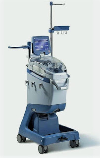
Cell Saver Machine


Definition:

-Cell (blood) salvage is a process in which a patient’s own (lost) blood is collected, processed, and transfused back (‘Autologous’ blood transfusion), which is done by a cell saver machine.

Principle:

1-Collection of blood: blood is suctioned from the operative field, and then heparinized saline is added, filtered, and centrifuged to separate RBCs which are then washed.

2-Washing of RBCs: across a semi-permeable membrane to filter out free Hb, plasma, WBCs, and platelets.

3-Re-transfusion: Washed RBCs are then suspended in saline (to achieve Hct of 60-70%) and transfused within 6 hours.

Advantages:

-Reduce or eliminate the need for ‘allogenic’ transfusion.

-‘Allogenic’ blood transfusion has been associated with an increased risk of postoperative infection, acute lung injury, perioperative MI, low CO HF, and tumor recurrence.

Indications:

-Anticipated blood loss >1L or >20% of estimated blood volume

-Patients with low Hb, multiple RBCs alloantibodies, rare blood group, and patient refusal of ‘allogenic’ blood transfusion

-Obstetrics: Controversial due to potential risk of amniotic fluid embolism. However; cell salvage with a Leucocyte-depletion filter (LDF) is considered safe

-Orthopedics: Reduce ‘allogenic’ transfusion & postoperative infection in arthroplasty

-Cardiac surgery: Leucocyte-depletion filter (LDF) use, reduce micro-emboli & lipid load of cell salvaged blood with an improvement of postoperative lung function.

-Vascular surgery

-Liver transplantation

-Jehovah’s Witness

Contraindications:

-Malignancy: due to risk of tumor dissemination

-Wound contamination: due to risk of systemic spread

-Old hemolyzed blood

-Use of collagen or hemostatic materials

-Obstetric surgery: due to risk of amniotic fluid embolism

-Ascites

Complications:

-Non-immune hemolysis: due to centrifugation

-Coagulopathy: due to large volumes of transfusion

-Citrate overdosage

-Air embolism

-Febrile non-hemolytic transfusion reaction

-Contamination: due to incomplete washing leading to contamination with drugs, activated leukocytes, cytokines, and microaggregates.

Dexmedetomidine

Dexmedetomidine

Mechanism of Action:

-It is an imidazole derivative and is a specific alpha-2 adrenoceptor agonist that acts via post-synaptic alpha-2 receptors primarily in the locus ceruleus to increase conductance through K+ channels.

Dexmedetomidine


Uses and Dose:

-Its main actions are sedation, anxiolysis, and analgesia

-It is a clear, colorless isotonic solution containing 100 g/ml of dexmedetomidine base and 9 mg/ml of sodium chloride in water. The solution is preservative-free and contains no additives.

-Dexmedetomidine can be administered intravenously, intramuscularly, and transdermally.

1. ICU Sedation:

-Used for sedation of initially intubated and mechanically ventilated patients in ICU.

-Loading: 1 mcg/kg IV over 10 minutes; loading dose may not be required for adults converted from other sedative therapy.

-Maintenance 0.2-0.7 mcg/kg/h. continuous IV infusion; not to exceed 24 h.

-The duration of use should not exceed 24 hours.

-Dexmedetomidine has been infused in mechanically ventilated patients before, during, and after extubation; it is not necessary to discontinue dexmedetomidine before extubation.

2. Procedural Sedation:

Indicated for sedation of non-intubated patients before and/or during surgical and other procedures.

Loading: 1 mcg/kg IV over 10 minutes.

Maintenance 0.6 mcg/kg/h. IV titrate to effect (usually 0.2-1 mcg/kg/h.).

3. Awake Fiberoptic Intubation:

Loading: 1 mcg/kg IV over 10 minutes.

Maintenance 0.7 mcg/kg/hr IV until endotracheal tube secured.

Dosage Modifications:

-Dose reduction may be required if co-administered with other concomitant anesthetics, sedatives, hypnotics, or opioids.

-Consider dose reduction in patients with hepatic impairment or aged ≥ 65 y.; clearance decreases with increasing severity of hepatic impairment.

-Renal impairment: No dosage adjustment required.

Pharmacokinetics

Distribution: It is 94% protein-bound in the plasma; the volume of distribution is 1.33 l/kg. The distribution half-life is 6 minutes.

Metabolism: The drug undergoes extensive hepatic metabolism to methyl and glucuronide conjugates.

Excretion: 95% of the metabolites are excreted in the urine. The elimination half-life is 2 hours, and the clearance is 39 l/hour.

Pharmacodynamics:

1. Cardiovascular System: It causes a predictable decrease in the mean arterial pressure and heart rate.

2. Respiratory System: It causes a slight increase in PaCO2 and a decrease in minute ventilation, with minimal change in the respiratory rate—these effects are not clinically significant.

3. Central Nervous System: The drug is sedative and anxiolytic—ventilated patients remain easily arousable and cooperative during treatment. Reversible memory impairment is an additional feature.

4. Metabolic / Other: It causes a decrease in plasma epinephrine and norepinephrine concentrations. It does not impair adrenal steroidogenesis when used in the short term.

Side Effects:

Hypotension, bradycardia, nausea, and a dry mouth are the most commonly reported side effects of the drug.

Anesthesia for Electroconvulsive Therapy

Anesthesia for Electroconvulsive Therapy

Anesthesia for Electroconvulsive Therapy


Principle:

-The exact mechanism of Electroconvulsive Therapy (ECT) is unknown. Electrical stimuli (electroconvulsive shock) are usually administered until a therapeutic generalized seizure is induced (30–60 sec. in duration).

-A good therapeutic effect is generally not achieved until a total of 400–700 seizures have been induced, in several sessions, over 2-3 weeks. Progressive short memory loss often occurs with an increasing number of treatments.

Physiological Effects:

-Seizure activity is characteristically associated with an “initial parasympathetic” discharge characterized by bradycardia and increased secretions. Marked bradycardia (<30 beats/min.) and even transient asystole (up to 6s) are occasionally seen.

-This is followed by “sustained sympathetic” discharge. Hypertension and tachycardia that follow are typically sustained for several minutes.

-Transient autonomic imbalance can produce arrhythmias and T-wave abnormalities on the ECG. Cerebral blood flow and ICP, intragastric pressure, and intraocular pressure all transiently increase.

Contraindications:

• Recent MI (<3 months)

• Recent stroke (usually <1 month)

• Intracranial mass or increased ICP from any cause

• More relative contraindications include:

- Angina

- Poorly controlled heart failure

- Significant pulmonary disease

-Bone fractures, Severe osteoporosis

- Pregnancy

- Glaucoma and retinal detachment.

Anesthetic Considerations:

-Amnesia is required only for a brief period (1–5 min) from when the NMB is given to when a therapeutic seizure has been successfully induced. The seizure itself usually results in a brief period of anterograde amnesia, somnolence, and often confusion. Consequently, only a short-acting induction agent is necessary.

-Increases in seizure threshold are often observed with each subsequent ECT.

-Most induction agents (Barbiturates, Benzodiazepines, and Propofol) have anticonvulsant properties, small doses must be used. The seizure threshold is increased and seizure duration is decreased by all of these agents.

--Sodium pentothal (2–4 mg/kg) was the first induction agent used, it raises the seizure threshold and decreases its duration.

--Methohexital (0.5-1.0 mg/kg): has been the induction agent of choice (gold standard) because it has very little effect on seizure duration and has a rapid onset and recovery profile. Unfortunately, methohexital is no longer available.

--Benzodiazepines: raise the seizure threshold and decrease its duration.

--Propofol (1–1.5 mg/kg): but higher doses reduce seizure duration.

--Etomidate (0.15-0.6 mg/kg): lacks anticonvulsant properties, increases seizure duration, and prolongs recovery.

--Ketamine (1.5-2 mg/kg): lacks anticonvulsant properties, and increases seizure duration, but is generally not used because it also increases the incidence of delayed awakening, nausea, and ataxia and is also associated with hallucinations during emergence.

-Short-acting opioids: are not given alone because they do not consistently produce amnesia.

-Sevoflurane (5%–8% for induction, followed by 1–2 MAC): is the only inhalational agent in widespread use for induction in ECT, with comparable effects to intravenous (IV) agents. It is preferred for patients not cooperative with IV access. It has the advantage of attenuating uterine contractions following ECT and is used in the third trimester of pregnancy.

-Induction agents in the descending order of seizure duration after their use are:

[Etomidate > Ketamine > Methohexital > Sevoflurane > Thiopental > Propofol]

-Induction agents in descending order of seizure threshold reducing property are:

[Etomidate > Ketamine > Methohexital > Thiopental > Propofol]

-Neuromuscular blockade: required from the time of electrical stimulation until the end of the seizure. A short-acting agent, such as succinylcholine (0.25–0.5 mg/kg), is most often selected.

-Ventilation: Controlled “mask ventilation” (with a backup plan of LMA if concerned about effective ventilation), is required until spontaneous respirations resume. As ECT is usually administered 3-times a week, repeated intubations may lead to airway trauma and edema. Hyperventilation can increase seizure duration and is routinely employed in some centers.

Monitoring:

-Routine monitoring should be as with the use of any other general anesthetic.

-Seizure activity is monitored by an unprocessed EEG. It can also be monitored in an isolated limb: a tourniquet is inflated around one arm before injection of succinylcholine, preventing entry of the NMB and allowing observation of convulsive motor activity in that arm.

Precautions:

-Rubber bite block: to avoid dental, tongue, and lips injury.

-Exaggerated parasympathetic effects: should be treated with atropine. In fact, premedication with glycopyrrolate is desirable both to prevent the profuse secretions associated with seizures and to attenuate bradycardia.

-Sympathetic manifestations: Nitroglycerin, Nifedipine, and α- and β-adrenergic blockers have all been employed successfully for control. High doses of β-adrenergic blockers (Esmolol, 200 mg), however, are reported to decrease seizure duration.

-Patients with pacemakers: may safely undergo ECT treatments, but a method to convert the pacemaker to a fixed mode, if necessary should be readily available.

Post-Tonsillectomy Bleeding

Post-Tonsillectomy Bleeding

Post-Tonsillectomy Bleeding


Incidence and Initial Management:

-The incidence of post-tonsillectomy bleeding ranges from 2% to 4%. It is more common in teenagers and young adults than in small children. The vast majority of postoperative bleeding occurs between days 5 and 10 when the eschar separates from the tonsil beds. In rare cases, bleeding may occur in the immediate postoperative period.

-Initial management is tailored to the degree of bleeding. The clot in the tonsillar bed should be suctioned, as spontaneous hemostasis is rare with the clot in place.

-Minimal bleeding may resolve with an ice water gargle or hydrogen peroxide gargle, and older children may tolerate the application of a cautery stick with silver nitrate while awake.

-Administration of antifibrinolytics as tranexamic acid.

-Significant bleeding in a young child, however, typically involves a return to the operating room for a thorough examination of the pharynx and electrocautery hemostasis.

Preoperative Assessment and Optimization:

1-Airway Assessment:

-Although it may be difficult to assess the airway in-depth in an agitated child, observation of the external anatomy and information about airway management from the prior anesthetic should provide sufficient information.

-Even though airway management was uncomplicated previously, it may be more difficult at this time because of postoperative edema and blood-obscuring visualization of the larynx.

2-Intravascular Volume Status:

-It is possible to underestimate the degree of blood loss, since much of it may have been swallowed. Heart rate, blood pressure, and, if possible, orthostatic testing will provide information regarding volume status and guide volume resuscitation.

-Assessing for the presence of tears, moist mucus membranes, skin turgor, and urine output will be helpful as well.

-Adequate IV access, with a large-bore intravenous catheter, should be obtained, if not already present.

-Intraosseous access should be entertained in the hypovolemic, hypotensive patient if IV access cannot be obtained in a timely fashion.

-Volume resuscitation should be initiated with non-glucose-containing isotonic fluids. If the patient is hypotensive, 20 mL/kg boluses of isotonic fluid should be administered until the blood pressure normalizes.

-Transfuse red blood cells if hemodynamically unstable and hematocrit is low.

-Once the bleeding has been controlled and the patient is normovolemic, maintenance fluids should be continued.

3-Laboratory Tests:

-Hematocrit, platelet count, electrolytes, and coagulation studies.

-A specimen should also be sent for serotyping and cross match, as transfusion is a very real possibility.

-Surgery should not be delayed waiting for test results, since bleeding will continue without surgical intervention

Intraoperative Management:

-Management of the airway is of major concern, particularly the ability to visualize the larynx in the presence of ongoing bleeding.

-In preparation for induction, the following should be prepared: multiple laryngoscope blades, a styletted cuffed ETT, and large bore suction.

-The otolaryngologist should be present and tracheostomy equipment immediately available should a surgical airway become necessary.

-The patient is considered to have a full stomach, even if they have not recently eaten, because of swallowed blood.

-A rapid sequence induction is usually performed. However, in the situation where there is significant concern about the airway, a smooth mask inhalation induction with cricoid pressure can be performed with the patient in the right lateral decubitus position with the head down (tonsillectomy position) with suction immediately available. The tonsillectomy position minimizes aspiration risk by promoting the pooling of blood in the oropharynx.

-The choice of IV induction agent will depend on the volume status. Ketamine or etomidate may be used if there is ongoing concern about volume status and hemodynamic stability. Alternatively, propofol may be used but in a decreased dose.

-Muscle relaxation can be achieved with either succinylcholine or rocuronium. However, the duration of action of rocuronium at the dose recommended for rapid sequence induction (1.2 mg/kg) will likely exceed the length of the procedure.

-Volume status should be continually assessed in the face of ongoing bleeding and managed appropriately with isotonic fluids.

-The hematocrit, degree of hemodynamic stability, and status of hemostasis will dictate the need for a blood transfusion.

-Replacement of coagulation factors is rarely necessary. However, if a previously undiagnosed coagulopathy is discovered, the appropriate therapy should be initiated and a hematology consult obtained.

Postoperative Management:

-Patients should be placed in the lateral decubitus position with the head down with supplemental oxygen for transport to the PACU.

-Analgesic and antiemetic medications should be ordered.