Educational Blog about Anesthesia, Intensive care and Pain management

Peripartum Cardiomyopathy (PPCM)

Anesthetic Management of Peripartum Cardiomyopathy (PPCM)


➧ It is a form of heart failure affecting females in their last months of pregnancy or early puerperium.

➧ The role of the anesthesiologist is important in the peri-operative and ICU.

Diagnostic Criteria for PPCM:

➧ Development of heart failure within the last month of pregnancy or 6 months postpartum.

➧ Absence of any identifiable cause for heart failure.

➧ Absence of any heart disease before the last month of pregnancy.

➧ Echo- Criteria of LV dysfunction:

-Ejection fraction < 45%.

-Left ventricular fractional shortening < 30%.

-Left ventricular end-diastolic dimension > 2.7 cm/m² BSA.

Risk factors:

-Black race, Family history.

-Advanced maternal age.

-Multiparity, Multiple gestations.

-Obesity, Malnutrition.

-Gestational HTN, Pre-eclampsia, C.S.

-Poor antenatal care, Breastfeeding.

-Alcohol, Cocaine, and Tobacco abuse.

Incidence:

-1 in 4,000 live births.

-This wide variation may be explained by the influence of genetic and environmental factors, as well as different reporting patterns and diagnostic criteria used.

Etiology:

1) Myocarditis

-It is not known whether it is an association or a cause.

-Only diagnosed by histological examination of endometrial biopsy.

-Also, the vasopressor therapy in PPCM may lead to Histological changes resembling myocarditis.

2) Viral Infection

-During pregnancy, there is a degree of depressed immunity that may lead to viral infection. Viral infection has been implicated as a cause of myocarditis that would lead to cardiomyopathy.

-On the other hand, it has been argued that viral cardiomyopathy should not be included as a cause of PPCM. But rather a separate entity.

3) Autoimmune Theory

-Studies hypothesized, that fetal cells may escape to the systemic circulation triggering an immune response.

-Higher rates of PPCM with twin pregnancies and its familial predisposition support this theory.

4) Inflammatory Cytokines

-In PPCM patients, higher concentrations of inflammatory cytokines like TNF α, CRP, and IL-6 were found. CRP levels correlated inversely with left ventricular ejection fraction (LVEF).

5) Selenium Deficiency

-Significantly low selenium concentrations in PPCM patients were found, still, this might be a mere incidental association rather than a cause.

6) Exaggerated Hemodynamic Response

-In pregnancy, there are physiologic changes in the CVS. It has been postulated that PPCM may be an exacerbation of this normal phenomenon.

7) Prolonged Tocolytic Therapy

-Usually, tocolysis causes tachycardia and vasodilation, so it may actually unmask existing heart disease rather than play an etiologic role.

Diagnosis:

-The symptoms & signs are the same as heart failure.

-You have to exclude other causes of heart failure as valvular and IHD.

Symptoms:

-Dyspnea on exertion, cough, orthopnea, and paroxysmal nocturnal dyspnea, resembling left-sided heart failure.

-Non-specific symptoms include palpitations, fatigue, malaise, and abdominal pain.

-Embolic manifestations may be present, as mural cardiac thrombi commonly occur. The patient may complain of chest pain, hemoptysis, and hemiplegia, rarely myocardial infarction may be the presentation due to coronary embolism.

Signs:

-Blood pressure may be normal/elevated/low.

-Tachycardia, Gallop rhythm.

-Engorged neck veins, Pedal edema

-Clinically, the heart may be normal or there may be mitral and/or tricuspid regurgitation with pulmonary crepitations.

Investigations:

➧ ECG: No specific findings.

➧ Chest x-ray: May show: Cardiomegaly, Pulmonary venous congestion.

➧ Echocardiography: It is the most important diagnostic tool, and assesses the severity and the prognosis of PPCM.

Echo findings are:

-Decreased LVEF and LVFS, Increased LVEDD.

-Dilatation of all cardiac chambers with subsequent functional mitral, tricuspid, pulmonary and aortic regurgitation.

➧ Dobutamine stress Echo: is a better prognostic tool than the ordinary Echo.

➧ TEE and Cardiac MRI: are better tools for detecting intramural thrombi than the ordinary Echo.

Complications:

1-Thromboembolism

-Thrombi often form in patients with LVEF < 35% and are associated with a mortality rate of 50%.

2-Arrhythmias

-All kinds of arrhythmias have been reported up to VT and heart arrest.

3-Organ failure

-Acute liver failure and hepatic coma due to passive liver congestion secondary to cardiac failure. Also, multiorgan failure may occur.

4-Obstetric & Perinatal complications

-There is an increased incidence of Abortion, Premature deliveries, IUGR, and Intrauterine fetal deaths.

Management:

A) Non-pharmacological measures:

-As with any heart failure, salt and water restriction (2-4 g/d., 2 L/d.).

-Once the severe symptoms are improved, modest exercise should be encouraged.

B) Pharmacological measures

-As with any heart failure: (Digoxin, Diuretics, VD, and Anticoagulation) are the mainstay.

-But we have to consider the safety of these drugs in pregnancy and lactation:

1-Digoxin

-It is a class C drug but presumed safe in low doses.

-In a pregnant female, the serum level should be monitored.

-Some studies claimed that digoxin for 6 m. decreases the risk of recurrence of PPCM.

2-Diuretics

-They are safe during pregnancy and lactation.

-Aim to reduce preload.

-Usually, loop diuretics are used and thiazides are used in milder cases. Spironolactone is very beneficial in heart failure but is better avoided during pregnancy.

-Should be used with caution not to induce dehydration and uterine hypoperfusion. Also, metabolic alkalosis may develop.

3-Vasodilators

-They reduce the preload and afterload in heart failure, and so increase the CO.

-Hydralazine and Nitrates are the VD of choice during pregnancy.

-ACEI and ARB are the mainstays in heart failure but they are class D, and contraindicated in pregnancy due to teratogenicity. So, they are considered after labor, but breastfeeding has to be discontinued.

4-Calcium Channel Modulators

-Calcium channel blocker: Although has –inotropic, but has been shown to improve the survival in cardiomyopathy patients. it also reduces the level of inflammatory cytokines so they would play an important in PPCM.

-Levosimendan: is especially valuable and used with success in PPCM. but again, breastfeeding should be avoided during its use.

5-Beta Blockers

-Like CCB, BB now has an important role in heart failure and they are not contraindicated in pregnancy, though associated with low birth weight.

-Both BB and ACEI have an additional role in immunosuppression and prevent remodeling and reduce ventricular dimensions.

6-Antiarrhythmic agents

-No antiarrhythmic agent is completely safe in pregnancy.

-Quinidine and Procainamide have a high safety profile, but treatment should always start in a hospital because of the high incidence of torsades de pointes.

-Amiodarone may cause: Hypothyroidism, Growth retardation, and Perinatal death, So it should be reserved for life-threatening arrhythmias only.

7-Anticoagulation therapy

-Anticoagulation therapy targets patients with LVEF < 35%, bedridden, Atrial fibrillation, Mural thrombi, Obese, or with a history of thromboembolism.

-The therapy may persist for as long as 6 w. in the Puerperium.

-Heparin is used in the antepartum and Heparin or Warfarin is used in the postpartum period as warfarin is contraindicated in the antepartum period due to its teratogenicity.

Obstetric management:

-Induction of delivery should be considered if pt. condition deteriorates despite maximal medical management.

-If the pt. is compensated, normal vaginal delivery is preferred.

-If the patient is severely decompensated or there are obstetric indications, C.S. should be done.

-In both cases, the pt. should be admitted to ICU for early detection of complications.

Anesthetic management:

A) Anesthesia for Vaginal Delivery:

-Controlled epidural a. under invasive monitoring is a safe and effective method.

-Sympathectomy induced by epidural leads to afterload and preload reduction that improves myocardial function in PPCM patients.

B) Anesthesia for Cesarean Section:

Both General anesthesia (GA) and Regional a. (RA) have been used.

1-Regional Anesthesia

-Single-shot spinal a. is not preferred, because of its rapid hemodynamic changes and hypotension.

-Epidural a. is used because of its better hemodynamic stability.

-Continuous spinal a., with its lower failure rates, faster onset, good muscle relaxation, less drug requirement, postoperative analgesia facilities, and better maintenance of hemodynamics has also been successfully applied.

-In severely compromised pt.: Local infiltration with Bilateral ilioinguinal blocks has been used.

2-General Anesthesia

-GA may be needed in emergency situations or when RA is contraindicated, particularly in the anticoagulated patient.

-Advantages: Airway control and ventilation, and it facilitates the use of TEE.

-Disadvantage: It can cause maternal and fetal cardiorespiratory depression, and the stress of rapid sequence induction on the decompensated heart could be dangerous. There is also an increased risk of LVF and pulmonary edema. GA does not provide thromboprophylaxis like RA.

-opioid-based anesthesia may be advantageous in compromised cardiac conditions, but carries a high risk of fatal respiratory depression.

-Monitoring: In mild cases, noninvasive monitoring can be used. in severely decompensated cases, the use of invasive monitoring is a must. This includes the use of an arterial line and may be a pulmonary artery catheter.

C) Postoperative management:

-All PPCM patients should be managed in an ICU as they are prone to develop LVF and pulmonary edema during this period. Also, to monitor the possible complications.

-Postoperative pain can be managed by RA or parenteral opioid-based techniques.

Prognosis:

➧ Poor prognosis criteria, the worst prognosis is found in patients with:

-Higher age and parity, Multiple gestations.

-Black race.

-Later onset of symptoms (> 2 w.) after delivery.

-Coexisting medical illness.

-Delay of initiation of medical treatment.

-Intracardiac thrombi, Conduction defects, Persistence of ventricular dysfunction > 6 m.

Risk of Recurrence in subsequent pregnancy:

-The highest risk of recurrence remains in patients with persistent cardiac dysfunction and the lowest risk is in those whose cardiac functions have been normalized, as evidenced by the dobutamine stress test.

INVOS™: In Vivo Optical Spectroscopy

INVOS™: In Vivo Optical Spectroscopy


Overview:

➧ INVOS™ system technology gives a noninvasive “window” to the body’s microvasculature; a direct and dynamic site of gas exchange that transports about half the body’s blood volume. 

➧ Measuring blood oxygenation in the microvasculature results in sensitive and site-specific insights on perfusion adequacy and multi-sensor monitoring gives data about perfusion distribution across the brain and body. 

➧ The non-invasive INVOS System reports the venous-weighted regional hemoglobin oxygen saturation (rSO₂) in the tissue under the sensor; reflecting the Hb bound O₂ remaining after tissues have taken what they need. Decreases in this venous reserve indicate increased ischemic risk and compromised tissue perfusion.

Clinical applications:

-Cerebral application: Brain area measurement

-Somatic application: Tissue area of measurement

Principle:

➧ The INVOS™ system utilizes near-infrared light at wavelengths that are absorbed by hemoglobin (730 and 810 nm). Light travels from the sensor’s light-emitting diode to either a proximal or distal detector, permitting separate data processing of shallow and deep optical signals.

➧ INVOS™ system’s ability to localize the area of measurement, called the spatial resolution, has been empirically validated in human subjects.

➧ Data from the scalp and surface tissue are subtracted and suppressed, reflecting rSO₂ in deeper tissues. This same concept applies to somatic monitoring.

➧ The result is continuous, real-time adequacy of perfusion data in up to four sites of your choice.

Clinical characteristics:

1-Noninvasive 

2-Continuous, real-time

3-Capillary (Venous and Arterial) sample

4-Measures the balance between O₂ supply and demand beneath the sensor

5-End organ oxygenation and perfusion

6-Requires neither pulsatility nor blood flow

Interpretation Values:

1-Cerebral: High blood flow, High O₂ extraction: 

➧ Typical rSO₂ range: 60-80%; assuming SpO₂ is > 90%.

➧ Common intervention trigger: rSO₂ < 50% or 20% change from rSO₂ baseline.

➧ Critical threshold: rSO₂ < 45% or 25% change from rSO₂ baseline.

2-Somatic: Variable blood flow, Lower O₂ extraction:

➧ Variances in the cerebral-somatic relationship may be indicative of pathology. 

➧ Watch for drops of 20% below patient baseline. 

Acute Kidney Injury Biomarkers

Acute Kidney Injury Biomarkers

I) Functional markers

1-Serum Creatinine (SCr):

➧ It is a degradation product of muscle cells and represents a surrogate for the efficiency of glomerular filtration.

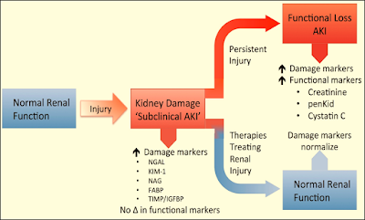
➧ It has poor predictive accuracy for renal injury, particularly, in the early stages of AKI.

➧ In the case of critical illness, SCr concentrations are subject to large fluctuations due to a patient’s induced dilutional volume status, the catabolic effects of critical illness, the likelihood of concentration decreases in septic conditions, and the increased tubular excretion with diminishing the renal function.

➧ Furthermore, after an injurious event, the rise in SCr is slow.

➧ Therefore, detection of the earliest evidence of AKI necessitates the use of other plasma or urinary biomarkers.

2-Plasma/Serum Cystatin C (CyC):

➧ It is a 13-kDa, non-glycosylated, cysteine protease inhibitor produced by all nucleated cells at a constant rate.

➧ In healthy subjects, plasma CyC (pCyC) is excreted through glomerular filtration and metabolized completely by the proximal tubules. There is no evident tubular secretion (not detectable in urine in healthy subjects).

➧ Several studies claim the superiority of pCyC against SCr to detect minor reductions in glomerular filtration rate (GFR).

➧ It is detected in plasma and urine 12-24 h. post-renal injury.

Confounding factors: older age, Gender, Weight, Height, Systemic inflammation, High levels of C-reactive protein, Malignancy, Thyroid disorders, immunosuppressive therapy, Glucocorticoid deficiency or excess, and Smoking.

3-Fractional Excretion of sodium (FENa):

➧ (FENa) measures the percent of filtered sodium that is excreted in the urine.

➧ This calculation is widely used to help differentiate prerenal disease (decreased renal perfusion) from acute tubular necrosis (ATN) as the cause of AKI.

➧ In pre-renal azotemia, the proximal tubules reabsorb filtered sodium resulting in a very low urine sodium concentration (<20 mmol/L) and FENa is <1%.

➧ In intrinsic AKI the urine sodium concentration is >40 mmol/L and FENa is >1%.

4- Proenkephalin A (Penkid):

➧ Penkid is a 5-kDa, stable breakdown product of enkephalins.

➧ It accumulates in the blood in settings of reduced GFR.

➧ It is associated with AKI and mortality in patients with sepsis and heart failure.

II) Low-molecular-weight proteins

1-Urine Cystatin C (uCyC):

➧ The urinary excretion of CyC (uCyC) specifically reflects tubular damage because systemically produced cystatin C is normally not found in urine.

2-Urine α1/β2 microglobulin:

III) Up-regulated proteins

1-Kidney Injury Molecule-1 (KIM-1) (Cytoprotection):

➧ It is a type I transmembrane glycoprotein with a cleavable ectodomain (90-kDa).

➧ It is localized in the apical membrane of dilated tubules in an acute and chronic injury.

➧ It is produced by proximal tubular cells after ischemic or nephrotoxic injury; no systemic source.

➧ KIM-1 plays a role in regeneration processes after epithelial injury and in the removal of dead cells in the tubular lumen through phagocytosis.

➧ A reduction in proteinuria with renin-angiotensin-aldosterone blockade is accompanied by a reduction in urinary KIM-1 excretion.

➧ It is detected in urine 12-24 h. after renal injury

Confounding factors: Renal cell carcinoma, Chronic proteinuria, Chronic kidney disease, Sickle cell nephropathy.

2-Neutrophil Gelatinase-Associated Lipocalin (NGAL) (also known as oncogene 24p3) (Iron binding):

➧ It is a 25-kDa glycoprotein produced by epithelial tissues throughout the body.

➧ It is a small protein linked to neutrophil gelatinase in specific leukocyte granules.

➧ It is also expressed in a variety of epithelial tissues associated with anti-microbial defense.

➧ NGAL’s composite molecule binds ferric siderophores, induces epithelial growth, has protective effects in ischemia, and is up-regulated by systemic bacterial infections.

➧ Plasma NGAL is excreted via glomerular filtration and undergoes complete reabsorption in healthy tubular cells. It is also produced in distal tubular segments.

➧ It is detected in plasma and urine 2-4 h. after AKI.

Confounding factors: Malignancy, Chronic kidney disease, Pancreatitis, COPD, Endometrial hyperplasia.

3-Liver Fatty Acid Binding Protein (L-FABP):

➧ They are small (15-kDa) cytoplasmic proteins (intracellular lipid chaperones) produced in tissues with active fatty acid metabolism (liver, intestine, pancreas, lung, nervous system, stomach, and proximal tubular cells).

➧ Their primary function is the facilitation of long-chain fatty acid transport, the regulation of gene expression, and the reduction of oxidative stress.

➧ Urinary (L-FABP) is undetectable in healthy control urine, which is explained by efficient proximal tubular internalization via megalin-mediated endocytosis.

➧ Under ischemic conditions, tubular L-FABP gene expression is induced. L-FABP is freely filtered in glomeruli and reabsorbed in proximal tubular cells; increasing urinary excretion after tubular cell damage.

➧ In renal disease, the proximal tubular re-absorption of L-FABP is reduced.

➧ Detected in plasma and urine 1 h. after ischemic tubular injury.

Confounding factors: Chronic kidney disease, Polycystic kidney disease, Liver disease, Sepsis.

4-Interleukin-18 (IL-18):

➧ It is 18-kDa pro-inflammatory cytokine, released from proximal tubular cells following injury.

➧ Detected in plasma and urine 6-24 h. after renal injury

Confounding factors: Inflammation, Heart failure, Sepsis.

5-Tissue Inhibitor of Metallo-Proteinases-2 (TIMP-2):

6-Insulin-like Growth Factor Binding Protein-7 (IGFBP-7):

➧ They are cell cycle arrest proteins that have been suggested as early indicators of AKI.

➧ In particular, urinary (TIMP-2) and (IGFBP-7) are biomarkers of the G1 renal tubular cell cycle arrest at the early phase of AKI.

➧ The product of the urinary concentrations of TIMP-2 and IGFBP-7 (urinary [TIMP-2] × [IGFBP-7]) is a promising biomarker for the early prediction of AKI.

IV) Tubular enzymes

1-Alpha-Glutathione-s-Transferase (α-GST):

2-Pi-Glutathione-s-Transferase (Ï€-GST):

➧ (α-GST) and (Ï€-GST) are 47-to 51-kDa cytoplasmic enzymes.

➧ They are both members of a multigene family of detoxification enzymes present in many organs including the kidney.

➧ Distribution across the entire nephron of structurally and functionally distinct isoforms has been demonstrated.

➧ In urine, these enzymes are normally not present.

➧ After the injury, α-GST is primarily detected in the proximal cells, whereas Ï€-GST is observed in the distal parts.

➧ They are detected in urine 12 h. after AKI.

3-Gamma-Glutamyl Transpeptidase (GGT):

4-Alkaline Phosphatase (AP):

5-Alanine Amino-Peptidase (AAP):

➧ They are tubular brush border enzymes.

➧ They are released into the urine when there has been significant damage to the brush border membrane with loss of the microvillus structures.

6-N-Acetyl-β-D-Glucosaminidase (NAG):

➧ N-Acetyl-β-D-Glucosaminidase (NAG) is a lysosomal enzyme (>130-kDa) that is localized in the renal tubules.

➧ It precludes glomerular filtration (due to large MW), implying that urinary elevations have a tubular origin.

➧ Increased activity suggests injury to its cells but may also reflect increased lysosomal activity without cell disruption.

➧ NAG catalyzes the hydrolysis of terminal glucose residues in glycoproteins.

➧ It is detected in plasma and urine 12 h. after AKI

Confounding factors: Diabetic nephropathy.

V) Others

1-Retinol Binding Protein (RBP):

➧ It is 21-kDa single-chain glycoprotein; a specific carrier for retinol in the blood (delivers retinol from the liver to peripheral tissues).

➧ It is totally filtered by the glomeruli and reabsorbed but not secreted by proximal tubules; a minor decrease in tubular function leads to the excretion of RBP in urine.

➧ It is detected in plasma and urine

Confounding factors: Type II DM, Obesity, Acute critical illness.

2-Hepcidin:

➧ It is a 2.78-kDa peptide hormone predominantly produced in hepatocytes; some production in the kidney, heart, and brain.

➧ It is freely filtered with significant tubular uptake and catabolism (fractional excretion 2%).

➧ It is detected in plasma and urine after AKI.

Confounding factors: Systemic inflammation.

3-Hepatocyte Growth Factor (HGF):

➧ It is overexpressed after AKI.

➧ It is a marker linked to renal tubular epithelial cell regeneration.

4-Netrin-1:

➧ It is a laminin-related molecule, minimally expressed in proximal tubular epithelial cells of normal kidneys.

➧ It is highly expressed in injured proximal tubules.

➧ It is detected in urine after AKI.

5-Monocyte Chemo-attractant Peptide-1 (MCP-1):

➧ It is a peptide expressed in renal mesangial cells and podocytes.

➧ It is detected in urine after AKI.

Confounding factors: Variety of primary renal diseases.

6-Calprotectin:

➧ The calcium-binding complex of two proteins of the S100 group (S100A8/ S100A9).

➧ Derived from neutrophils and monocytes.

➧ Acts as an activator of the innate immune system.

➧ It is a measure of local inflammatory activity. It is detected in urine in intrinsic AKI.

Confounding factors: Inflammatory bowel disease, Urinary tract infection, Probably CKD.

7-MicroRNA:

➧ MicroRNAs are short, non-protein-coding RNA molecules between 19 and 25 nucleotides in length.

➧ They are epigenetic regulators of gene expression at the post-transcriptional level in response to kidney injury through messenger RNA (mRNA) signal repression.

➧ As key regulators of homeostasis, their dysregulation underlies several morbidities including kidney disease.

➧ MicroRNAs are used as diagnostic and prognostic biomarkers in AKI.

8- Chitinase-3-like protein 1 (CHI3L1) (YKL-40, HC-gp39):

➧ YKL-40 is a 40-kDa heparin- and chitin-binding glycoprotein also known as Human Cartilage glycoprotein 39 (HC-gp39), 38-kDa heparin-binding glycoprotein or chitinase-3-like protein 1 (CHI3L1).

➧ The abbreviation YKL-40 is based on the one-letter code for the first three N-terminal amino acids, tyrosine (Y), lysine (K), and leucine (L), and the apparent molecular weight of YKL-40.

➧ It plays an important role in AKI and repair.


Acute Kidney Injury Biomarkers

Acute Kidney Injury Biomarkers

Acute Kidney Injury Biomarkers


Bronchoscopy in ICU

Bronchoscopy in ICU


Indications:

A) Diagnostic:

1-Investigation of an infectious process:

➧ Bronchoscopy and broncho-alveolar lavage (BAL) are commonly used for the identification of pathogens in the lower airways in the following circumstances: 

a) When it is not clear whether the presence of organisms represents infection or colonization. 

b) When the infection is caused by organisms that show a predilection for the peripheral airways (e.g. Pneumocystis jirovesii). 

c) When the infection is confined in a particular area of the lung (tuberculous and non-tuberculous mycobacteria as well as several fungal and opportunistic organisms).

2-Abnormal breath sounds:

➧ Abnormal breath sounds include: stridor, wheezes, or a combination of both and they are the most common indication of FOB in the NICU.

➧ In neonates, abnormal breath sounds are due to congenital abnormalities of the upper or lower airways.

➧ In older infants and children the causes are often iatrogenic (e.g. subglottic stenosis due to prolonged intubation, injury of the vocal cords, or recurrent laryngeal nerve causing paresis or paralysis).

3-Evaluation of the nature of abnormalities:

➧ Patients in the ICU often present with radiographic findings and/or symptoms of unclear etiology, e.g.: an airway filled with secretions can look radiographically identical to a completely compressed airway.

4-Others:

➧ Pneumonia, Trauma, Inhalation injury and burns, Tracheoesophageal fistula.

B) Therapeutic:

1-Placement of the endotracheal tube:

➧ The use of FOB for the placement of the endotracheal tube is reserved for cases in which high precision is required.

➧ e.g. placement of the ETT just above the carina in patients with very severe tracheomalacia; or selective intubation of one lung), or when congenital anatomical abnormalities or injuries preclude the proper opening of the jaw for direct laryngoscopy.

2-Placement of double-lumen ETT and confirmation of position

3-Extubation over FOB

4-Persistent or recurrent atelectasis

➧ Atelectasis is a major cause of clinical deterioration and/or of delay in the patients’ recovery, resulting from many causes such as; mucous plug, compression of the airways, alveolar destruction, and collapse.

5-Foreign body removal

6-Strictures and stenosis

7-Hemoptysis

Contraindications:

1-Full stomach


3-Severe pulmonary hypertension

4-Tuberculosis

5-Acute myocardial infarction or unstable angina

6-Coagulopathy

Complications:

1-Increase HR, Bl. P, ICP, IOP

2-Laryngospasm and bronchospasm

3-Hypoxemia

4-Arrhythmia

5-Bleeding

6-Post-bronchoscopy fever

7-Pulmonary infiltrate

Diabetic Ketoacidosis (DKA)

Diabetic Ketoacidosis (DKA)


Physiology:

Hyperglycemia:

➧ Increased hepatic production of glucose.

➧ Diminished glucose uptake by peripheral tissues.

➧ Insulinopenia / Hyperglucagonemia.

Ketoacidemia:

➧ The ketoacid is acetoacetic acid. The byproduct is acetone. The non-keto-acid is beta-hydroxybutyric acid.

➧ Increased lipolysis and hepatic ketogenesis

➧ Reduced ketolysis by insulin-deficient peripheral tissues.

Fluid and Electrolyte Depletion:

➧ Osmotic diuresis and dehydration due to hyperglycemia.

➧ On average, water deficit is about 5L, sodium 500 mmol, potassium 400 mmol, and chloride 400 mmol.

General Considerations:

➤ Initial presentation of Type I DM (Can also occur in Type II DM).

➤ Increased insulin requirements in Type I DM (Infection, Trauma, Myocardial infarction, Surgery).

➤ Mortality is 5% in patients under 40 y. Up to 20% of the elderly.

➤ Estimates of 5-8 episodes per 1000 at-risk diabetics annually.

➤ One of the more common serious complications of insulin pump users – occurs 1 per 80 months of treatment. Typically due to unrecognized pump failure.

Essentials of Diagnosis:


➧ Acidosis with pH < 7.3.

➧ Serum bicarbonate < 15 .mEq/L.

➧ The serum is positive for ketones.

➧ Elevated anion gap (variable, may occur without gap).

➧ Hyperglycemia > 250 mg/dL (no correlation between the severity of hyperglycemia and severity of ketoacidosis).

Clinical picture:

Symptoms:

➧ Early: Polyuria, Polydipsia, Fatigue, N/V.

➧ Late: Stupor – Coma.

Signs:

➧ Rapid, Deep Breathing.

➧ Fruity breath odor of acetone.

➧ Tachycardia, Hypotension, mild Hypothermia.

➧ Abdominal Pain and Tenderness.

Laboratory Findings:

➧ Glycosuria 4+, Ketonuria.

➧ Hyperglycemia, Ketonemia, Low arterial blood pH, and Low plasma bicarbonate.

➧ Elevated serum potassium (despite total body potassium depletion).

➧ Elevated serum amylase (not specific for pancreatitis in this setting, use lipase).

➧ Leukocytosis.

➧ If hyperthermic, likely due to infection since pts with DKA are hypothermic if uninfected.

Management of DKA:

Insulin Replacement:

➧ Regular Insulin IV bolus 0.1-0.2 units/kg to ‘prime’ insulin receptors.

➧ Regular Insulin infusion at 0.1 units/kg/h.

➧ Then replaced with SC regular insulin when hyperglycemia and ketoacidosis are controlled.

➧ Then oral intake + SC intermediate-acting insulin.

Fluid Replacement:

➧ The typical deficit is 4-5 L.

➧ Initially, NS 1 L/h. x 2 h., then 0.5 L/h. x 1-2 h., then 200-300 mL/h. till correction.

➧ Switch to ½ NS if serum Na > 150 mEq/L. 

➧ Add D5W if the glucose falls below 250 mg/dL, to maintain serum glucose 250-300 mg/dL to prevent hypoglycemia and cerebral edema.

Sodium Bicarbonate:

➧ 50 mmol

➧ Clinical benefit has not been demonstrated.

➧ Use to correct pH < 7, target pH of 7-7.2.

Potassium:

➧ 10-30 mEq/h. replacement to be started during the second or third hour of treatment.

Phosphate:

➧ Replete hypophosphatemia of < 1 mg/dL.

➧ 15 mmol K or Na phosphate in 100 mL saline.

➧ Replete slowly (3-4 mmol/h.) to avoid hypocalcemic tetany.

Treatment of Associated Infection:

➧ Antibiotics: as indicated.

➧ Cholecystitis and pyelonephritis may be particularly severe in these patients.

Read more ☛ Hypoglycemic Coma

Hypoglycemic Coma

Hypoglycemic Coma


Hypoglycemia in Type I DM:

➧ Common in patients intensively controlled with insulin.

➧ Asymptomatic blood glucose levels of < 50 mg/dL occur daily in up to 56% of patients.

➧ Symptomatic hypoglycemia occurs 2X/week on average.

Severe Hypoglycemia:

➧ An episode requires intervention by another person for the patient to recover function.

Causes of Hypoglycemia in Diabetes:

➧ Delayed, reduced, or missed CHO intake.

➧ Increased glucose utilization (exercise).

➧ Decreased insulin clearance (renal failure).

➧ Alcohol -inhibits hepatic gluconeogenesis.

Adrenal insufficiency or glucocorticoid dosage reduction.

Clinical picture:

Adrenergic:

➧ Tremor, anxiety, palpitations, hunger.

Neuroglycopenic:

➧ Dizziness, decreased concentration, blurred vision, tingling, lethargy.

Severe Hypoglycemia in Intensively Controlled Type I DM:

➧ Up to 25% yearly incidence.

➧ Disabling cognitive effects may take hours to fully resolve.

➧ May lead to seizures, and rarely, permanent neurological deficits.

➧ Estimated to be a causative factor in 4% of deaths.

Hypoglycemia Unawareness:

➧ Loss of autonomic warning symptoms of hypoglycemia.

➧ Occurs in 25-50% of patients with type I DM.

➧ Patients are no longer prompted to eat.

➧ Results in a 7X increased frequency of severe hypoglycemia.

Defective Glucose Counter-regulation in Type I DM:

➧ Reduced or absent glucagon response is common after 2-4 years.

➧ Deficient epinephrine response is common after 5-10 years.

➧ Results in a 25X increased frequency of severe hypoglycemia.

Hypoglycemia Unawareness and Defective Glucose Counter-regulation:

➧ Reversible by short-term avoidance of hypoglycemia.

Reduction of Hypoglycemia in Type I DM:

➧ Identify patients at increased risk:

- History of severe hypoglycemia.

- History of hypoglycemia unawareness.

- Normal or near-normal glycohemoglobin levels.

➧ Raise glycemic targets in the short term to regain symptom recognition.

➧ Education of patients and family members to recognize and treat hypoglycemia.

➧ Have unaware patients test blood glucose before performing a critical task (driving).

➧ Patients should have rapid-acting carbohydrates available at all times.

➧ Apply principles of intensive insulin therapy:

- Frequent home glucose monitoring.

- Flexible insulin regimens with dosage adjustments based on meal size, monitored blood glucose levels, and anticipated exercise.

➧ Replace insulin more physiologically:

- Multiple insulin injections.

- New ultra-short-acting insulin analogs: lispro, aspart, glulisine.

- Long-acting insulin analogs: glargine, detemir.

- Insulin pumps.

Subcutaneous, Continuous Glucose Monitors:

➧ Now available with alarms for high and low glucose readings.

➧ Useful for catching periods of hypoglycemia (especially overnight) of which patients are unaware.

➧ Shown to reduce the incidence of hypoglycemia in type I DM patients with prior severe hypoglycemia.

Management of Hypoglycemic Coma:

-If delayed, can cause permanent neurologic damage.

➧ 50% Dextrose in water: 50 ml IV over 3-5 min. followed by 5% dextrose in a water infusion.

➧ Glucagon: 0.5-1 mg IM/SC.

- Mobilizes hepatic glycogen stores.

➧ Hydrocortisone: for adrenal insufficiency.

➧ Hospitalize: those on sulfonylureas for 24 h.