Educational Blog about Anesthesia, Intensive care and Pain management

Down's Syndrome

Anesthetic Management of Down's Syndrome


➧ This well-known syndrome, with characteristic morphological features and mental retardation, results from the chromosomal abnormality, trisomy 21.

➧ Anesthetic risk is increased in these children. Indeed, the mortality is increased at any stage of life, but improved medical and nursing care means that many more individuals are surviving into adulthood and may present for surgery.

➧ Between 60 and 70% of patients now survive beyond 10 years of age.

Preoperative abnormalities:

1. Cardiac abnormalities: occur in 50–60% of patients and are usually responsible for the initial mortality in infancy. The commonest lesions are septal defects, Fallot’s tetralogy, and patent ductus arteriosus. In adults, there is an increased risk of mitral valve prolapse and mitral and aortic valve regurgitation.

2. Immune system defect: results in an increased incidence of infection. Granulocyte abnormalities decreased adrenal responses, and defects in cell-mediated immunity have all been identified. There is an increased incidence of lymphomas and leukemias.

3. Skeletal abnormalities: atlantoaxial instability was recognized as being a problem, at a time when these children were encouraged to participate in gymnastics. Down’s children may have C1–C2 articulation abnormalities, subluxation, and odontoid peg abnormalities. It may occur in association with either medical procedures or physical activity. The cause of instability may be due to: poor muscle tone, ligamentous laxity, and abnormal development of the odontoid peg.

4. Biochemical abnormalities: involve the metabolism of serotonin, catecholamines, and amino acids.

5. Thyroid hypofunction: is common in both adults and children, although hyperthyroidism can sometimes occur. A child with Down’s syndrome had a thyrotoxic crisis that mimicked malignant hyperthermia.

6. Sleep-induced ventilatory dysfunction: has been reported.

7. Institutionalized Down’s patients have an increased incidence of hepatitis B antigen.

8. Autonomic dysfunction: in particular increased sympathetic function and decreased vagal activity, result from brainstem abnormalities.

Anesthetic problems:

1. Cervical spine abnormalities: increase the risk of dislocation of certain cervical vertebrae on intubation, or when the patient is paralyzed with muscle relaxants with risk of atlantoaxial subluxation and spinal cord compression. Cervical spine screening has to be carried out, and precautions taken during intubation. 

2. The larynx is often underdeveloped and smaller tracheal tube size is required than would be anticipated for the age of the patient. The adult larynx may only accept a size 6-mm tube.

3. Airway and intubation difficulties sometimes occur, from a combination of anatomical features. These include a large tongue, a small mandible and maxilla, a narrow nasopharynx, and irregular teeth. Even in the absence of teeth, intubation is made more difficult by excessive pharyngeal tissue.

4. Postoperative stridor after prolonged nasal intubation. Congenital subglottic stenosis occurs occasionally.

5. Obstructive sleep apnea is common in Down’s syndrome. Compared with normal children they had an increased incidence of stridor and chest wall recession, lower baseline oxygen saturations, and a greater number of episodes of desaturation to 90% or less. Chronic episodes of hypoxia and hypercarbia may lead to pulmonary hypertension and congestive heart failure. Airway patency depends upon both the anatomical structure of the upper respiratory tract and the normal functioning of the pharyngeal muscles. Abnormalities of either or both may occur.

6. Upper airway obstruction, because it has multiple causes, is not necessarily resolved with surgical treatment. Young patients with more severe symptoms often had multiple sites of obstruction and a high incidence of cardiac disease.

7. Problems of the associated cardiac disease, which in later life may lead to pulmonary hypertension.

8. A high incidence of atelectasis and pulmonary edema after surgery for congenital heart disease. Those with Down’s syndrome and ventricular septal defects were predisposed to pulmonary vascular obstruction.

9. Posterior arthrodesis of the upper cervical spine carries a high complication rate. Problems included infection and wound dehiscence, instability at a lower level, neurological sequelae, and postoperative death.

Management:

1. Lateral cervical X-rays are required, in full flexion and extension positions, to detect atlantoaxial instability. This may show as an increase in the distance between the posterior surface of the anterior arch of the atlas, and the anterior surface of the odontoid process. Patients with an atlanto-odontoid interval of 4.5–6.0 mm were asymptomatic, but those in whom the distance exceeded 7 mm had neurological signs. If instability is present, great care should be taken to immobilize the neck during intubation and muscle relaxation. These changes do not appear to progress with time. 

2. If a significant cardiac disease is present, management must be appropriate to the lesion, and endocarditis prophylaxis is given as recommended.

3. A tracheal tube should be used that is 1–2 sizes smaller than would be expected from the patient’s age.

4. If prolonged nasotracheal intubation is required, steroids should be given before extubation. The child should receive humidification and be observed carefully for signs of stridor.

5. Close observation is required in the perioperative period, to detect episodes of obstructive apnea. A pulse oximeter is useful.

6. Loss of locomotor skills or disturbances of gait after surgery or acute trauma should alert staff to the possibility of subluxation and cord compression. In the event of this, an urgent neurological opinion should be sought. However, in the absence of neurological signs, non-operative management has been advised, because of the high complication rate after surgery.

7. In patients with adenotonsillar hypertrophy, surgery may improve obstruction. However, close monitoring and oxygen therapy are important for the first postoperative night.

Propofol Related Infusion Syndrome (PRIS)

Propofol Related Infusion Syndrome (PRIS)


➧ It is a rare syndrome that affects patients undergoing long-term treatment with high doses of the anesthetic and sedative drug propofol. 

➧ It is associated with high doses and long-term use of propofol (>4 mg/kg/hr for more than 24 hours). It occurs more commonly in children, and critically ill patients receiving catecholamines and glucocorticoids are at high risk.

Clinical Picture:

➧ Arrhythmias 

➧ Progressive Myocardial Failure 

➧ Cardiovascular Collapse 

➧ Lipemia 

➧ Hypertriglyceridemia 

➧ Acute Renal Failure 

➧ Rhabdomyolysis 

➧ Metabolic acidosis 

➧ Hyperkalemia 

➧ Hepatomegaly 

➧ Green urine (phenol metabolites), (Figure 1) 

➧ It is often fatal 


Green Urine
Figure 1: Green Urine

Laboratory Results:

➧ Elevated serum lactate 

➧ Elevated CPK 

➧ Myoglobinuria 

➧ Hyperkalemia 

➧ Hypertriglyceridemia

ECG changes:

➧ ST elevation in precordial leads V1 to V3

Predisposing factors for developing PRIS:

➧ Propofol dose >4 mg/kg/h 

➧ Propofol infusion >48 h 

➧ Presence of “triggering factor” (i.e. catecholamine infusion or corticosteroids) 

➧ Inadequate delivery of carbohydrate 

➧ Critical Illness 

➧ Severe Cerebral Injury 

➧ Sepsis 

➧ Pancreatitis 

➧ Trauma

Prevention:

➧ Avoid high propofol doses and minimize the duration of infusion in high-risk patients 

➧ Avoid lipid overload by assessing all sources of fat calories (e.g., parenteral nutrition, enteral nutrition). 

➧ Monitoring of serum triglycerides in any patient receiving propofol in doses >4 mg/kg/h or >48 is highly recommended. 

➧ Assure adequate provision of carbohydrates. 

➧ Depletion of carbohydrate stores can promote mobilization of fat stores and increase lipid metabolism. This, in turn, increases circulating fatty acid load and may predispose patients to PRIS. 

➧ Theoretically, it is, therefore, possible that early adequate carbohydrate intake may prevent PRIS by preventing the switch to fat metabolism. 

➧ There is some suggestion that providing a carbohydrate intake of 6–8 mg/kg/min. can suppress fat metabolism and thus prevent PRIS.

Treatment: (Supportive) 

➧ Early recognition of the syndrome and discontinuation of the propofol infusion reduces morbidity and mortality. 

➧ Once a patient presents with symptoms compatible with PRIS, propofol infusion should be discontinued promptly and an alternative sedative agent should be initiated. 

➧ Cardiovascular support by the combination of vasopressors and inotropes. 

➧ Cardiac pacing should be considered. 

➧ Hemodialysis or hemofiltration to decrease the plasma concentrations of circulating metabolic acids and lipids. 

➧ Extracorporeal membrane oxygenation (ECMO) for combined respiratory and circulatory support. 

Central Anticholinergic Syndrome

Central Anticholinergic Syndrome

➧ Many of the drugs used in anesthesia and intensive care may cause blockage of the central cholinergic neurotransmission.

➧ Acetylcholine is of significance in the modulation of the interaction among most other central transmitters.

Causes:

1- Overdose of anticholinergic drugs: e.g. Atropine, scopolamine, glycopyrrolate

2- Overdose of drugs that possess an anticholinergic activity:
e.g. Antipsychotics, tricyclic antidepressants, antiparkinsonian drugs

3- It May be induced by opiates, benzodiazepines, phenothiazines, butyrophenones, ketamine, etomidate, propofol, nitrous oxide, and halogenated inhalation anesthetics as well as by H2-blocking agents such as cimetidine.

Clinical Picture:

➧ The clinical picture of the central cholinergic blockade, known as the central anticholinergic syndrome (CAS), is identical to the central symptoms of atropine intoxication.

➧ This behavior consists of agitation including seizures, restlessness, hallucinations, disorientation, or signs of depression such as stupor, coma, and respiratory depression.

Systemic:

➧ Tachycardia: Increased heart rate

➧ Ataxia: loss of coordination 

➧ Dry, sore throat, xerostomia, or dry mouth with a possible acceleration of dental caries 

➧ Cessation of perspiration → Increased body temperature 

➧ Mydriasis: Pupil dilation; consequent sensitivity to bright light (photophobia) 

➧ Cycloplegia: Loss of accommodation (loss of focusing ability, blurred vision) 

➧ Diplopia: Double-vision 

➧ Increased intraocular pressure; dangerous for people with narrow-angle glaucoma

➧ Urinary retention, Diminished bowel movement, and sometimes ileus

➧ Shaking

Central nervous system:

Resemble those associated with delirium, and may include:

➧ Mental confusion (brain fog), Disorientation, Agitation, Irritability, Unusual sensitivity to sudden sounds 

➧ Euphoria or dysphoria 

➧ Memory problems, Inability to concentrate

➧ Illogical thinking, Wandering thoughts; inability to sustain a train of thought 

➧ Incoherent speech 

➧ Respiratory depression 

➧ Wakeful myoclonic jerking 

➧ Photophobia 

➧ Visual disturbances: Periodic flashes of light, changes in the visual field, Visual snow, Restricted or "tunnel vision" 

➧ Visual, auditory, or other sensory hallucinations: 

-Warping or waving of surfaces and edges 

-Textured surfaces 

-"Dancing" lines; "spiders", insects

-Lifelike objects indistinguishable from reality 

-The hallucinated presence of people not actually there 

➧ Rarely: seizures, coma, and death 

➧ Orthostatic hypotension (sudden dropping of systolic blood pressure when standing up suddenly) and significantly increased risk of falls in the elderly population.

Treatment:

Central anticholinergic syndrome is completely reversible and subsides once all of the causative agents have been excreted.

Physostigmine:

➧ It is one of only a few drugs that can be used as an antidote for anticholinergic poisoning because it crosses BBB.

- Dose: 0.01-0.03 mg/kg IV, repeated after 15-30 min. If necessary. 

- Duration of action: about 30-60 min after intravenous IV injection.

- Side effects: nausea and vomiting, abdominal cramps, bradycardia, and hypotension

- Physostigmine was found to increase the risk of cardiac toxicity.

Nicotine:

➧ It counteracts anticholinergics by binding to the nicotinic acetylcholine receptors and activating them.

Caffeine:

➧ It is an adenosine receptor antagonist, that counteracts the anticholinergic symptoms by reducing sedation and increasing acetylcholine activity, thereby causing alertness and arousal.

➧ Caffeine can also inhibit acetylcholinesterase non-competitively.

Fospropofol disodium (Lusedra®)

Fospropofol disodium (Lusedra®)


Chemistry:

➧ The chemical nature of propofol is 2,6-diisopropyl phenol. When a phosphate group is added to this molecule, it results in the formation of water-soluble propofol that does not contain lipids, egg products, or preservatives, thereby eliminating the allergic, bacterial infections and hyperlipidemic concerns associated with propofol. 

➧ The two phosphorylated propofol prodrugs were named propofol phosphate and phosphonooxymethyl propofol. 

➧ Substitution of hydroxyl by charged phosphate group introduces electronegativity which allows fospropofol to dissolve readily in water, hence does not cross the lipid membrane. The sodium salt of fospropofol is commonly used (2,6-diisopropyl phenoxy methyl phosphate disodium salt).

Mechanism of Action:

➧ Fospropofol is a water-soluble prodrug of propofol, a sedative/hypnotic/anesthetic drug. it is hydrolyzed by endothelial alkaline phosphatases to an active metabolite, propofol, formaldehyde, and phosphate, this process takes 15-20 min. leading to slow onset of action (4-13 min.). Its mechanism of action is uncertain, but it is postulated that its primary effect may be potentiation of the GABA-A and glycine receptors, possibly by slowing the channel closing time.

Uses:

➧ Fospropofol is an intravenous sedative-hypnotic agent. It is currently approved for use in monitored anesthesia care sedation of adult patients undergoing diagnostic or therapeutic procedures such as endoscopy.

Formulation and dose:

➧ Fospropofol is supplied as an aqueous solution designed for intravenous injection. The recommended initial dose of the drug is as follows:

Standard Dosing Regimen:

➧ In adults aged 18 to <65 years, who are healthy or have mild systemic disease, an initial IV bolus of 6.5 mg/kg followed by supplemental IV doses of 1.6 mg/kg (25 % of initial dosage) as needed to achieve the desired level of sedation. 

➧ The dosage of fospropofol is limited by the lower and upper weights of 60 kg and 90 kg.

Modified Dosing Regimen:

➧ For adults>65 years of age or those with severe systemic disease, initial and supplemental IV dosages of 75 % of the standard dosing regimen.

Advantages:

1-Less pain at the site of IV administration. 

2-Less potential for hyperlipidemia with long-term administration. 

3-Less chance for bacteremia.

Side Effects:

➧ Adverse events associated with the use of fospropofol may include, but are not limited to, the following: 

1-Paresthesias 

2-Pruritus 

3-Hypotension 

4-Hypoxemia 

5-Nausea 

Magnesium Sulphate (MgSO₄)

Magnesium Sulphate (MgSO₄)


Magnesium Physiology:

➧ Magnesium is the fourth most abundant cation in the body and is the second most abundant intracellular cation in the human body (potassium being the first).

➧ It has numerous physiological activities including activation of enzymes involved in energy metabolism, protein synthesis, regulation of vasomotor tone, neurotransmission, and signaling.

➧ It serves as a cofactor for more than 300 enzyme reactions that involve adenosine triphosphate (ATP). One of the magnesium-dependent enzyme systems is the membrane pump that generates the electrical gradient across cell membranes. As a result, magnesium plays an important role in the activity of electrically excitable tissues.

➧ Magnesium regulates the movement of calcium into smooth muscle cells, which gives it a pivotal role in the maintenance of cardiac contractile strength and peripheral vascular tone.

➧ Magnesium is required for the transformation of thiamine into thiamine pyrophosphate, so a magnesium deficiency can promote thiamine deficiency in the face of adequate thiamine intake. For this reason, the magnesium status should be monitored periodically in patients receiving daily thiamine supplements.

➧ Magnesium also has antinociceptive effects in animal and human pain models. Magnesium antinociceptive effects appear to be relevant not only to chronic pain but also, to the duration and intensity of postoperative pain. These effects are primarily based on physiological calcium antagonism, that is voltage-dependent regulation of calcium influx into the cell, and noncompetitive antagonism of NMDA receptors which produces a reduction of NMDA-induced currents.

Magnesium Balance:

➧ The average-size adult contains approximately 24 g (1 mole, or 2000 mEq) of magnesium. 

➧ Half of the total body content of magnesium is found in bone.

➧ The muscle and liver are the soft tissues that contain the greatest amount of magnesium.

➧ Thirty percent of extracellular magnesium circulates bound to protein. Therefore, albumin concentration must be known to interpret total magnesium levels.

Corrected Serum Mg = Mg x 0.42 + 0.05 (4 - albumin in g/dL) 

➧ Less than 1% of magnesium is located in plasma. This lack of representation in the plasma limits the value of the plasma magnesium concentration as an index of total body magnesium stores. 

➧ This is particularly true in patients with magnesium deficiency, in whom serum magnesium levels can be normal in the face of total body magnesium depletion.

➧ The serum is favored over plasma for magnesium assays because the anticoagulant used for plasma samples can be contaminated with citrate or other anions that bind magnesium. The normal range for serum magnesium depends on the daily magnesium intake, which varies according to geographic region.

➧ Only 67% of the magnesium in plasma is in the ionized (active) form, and the remaining 33% is either bound to plasma proteins (19% of the total) or chelated with divalent anions such as phosphate and sulphate (14% of the total).

➧ The daily oral intake is 8–20 mmol (40% of which is absorbed). Magnesium absorption occurs throughout the small intestine and is enhanced by 1,25(OH)₂D₃. Regulation of magnesium balance is mainly by the kidneys. Like calcium, magnesium is reabsorbed in the kidney tubules.

➧ When magnesium intake is deficient, the kidneys conserve magnesium and urinary magnesium excretion falls to negligible levels and the serum magnesium remains in the normal range. This illustrates the relative value of urinary magnesium over serum magnesium levels in the detection of magnesium deficiency.

➧ The normal range for healthy adults residing in the United States is shown in Table 1:

Magnesium Reference Ranges
Table 1: Magnesium Reference Ranges

Magnesium Sulphate Preparations:

➧ The standard intravenous preparation is magnesium sulphate (MgSO₄). Each gram of magnesium sulphate has 8 mEq (4 mmol) of elemental magnesium.

➧ Saline solutions should be used as the diluent for magnesium sulphate. Ringer's solutions should not be used because the calcium in Ringer's solutions will counteract the actions of the infused magnesium.

Uses of Magnesium Sulphate:

1- Prevention of eclampsia in women with pre-eclampsia and it is also a recommended treatment for established eclampsia.

2- Used to treat atrial fibrillation, to achieve both rate control and reversion to sinus rhythm in many settings, including post-cardiac surgery, and in the emergency department.

3- Intravenous magnesium can suppress digitalis-toxic arrhythmias, even when serum magnesium levels are normal. Intravenous magnesium can also abolish refractory arrhythmias (i.e., unresponsive to traditional antiarrhythmic agents) in the absence of hypomagnesemia. This effect may be due to a membrane-stabilizing effect of magnesium that is unrelated to magnesium repletion.

4- It May be beneficial for patients with acute severe asthma either given intravenously or nebulized. 

5- May also prevent delayed cerebral ischemia due to vasospasm in patients with subarachnoid hemorrhage. 

6- Used in hypomagnesemia and hypomagnesemia associated with cardiac arrhythmias. 

7- Magnesium sulphate when used systemically, has shown antinociceptive effects and has decreased postoperative opioid requirements. A limitation to the parenteral application of magnesium for modulation of antinociception via NMDA channel antagonism is insufficient blood-brain barrier penetration to achieve effective CSF concentrations. 

8- Intrathecally administered magnesium has antinociceptive effects in animals. Intrathecal magnesium, an NMDA antagonist, has been demonstrated to prolong analgesia without notable adverse effects in healthy parturients and in various surgical procedures like lower limb surgeries and in patients undergoing total abdominal hysterectomy.

Contraindications:

Hypocalcemia, heart block (risk of arrhythmias), and oliguria.

Interactions:

➧ Potentiates neuromuscular blockade (nondepolarizing/depolarizing). 

➧ Potentiates CNS effects of anesthetics, hypnotics, and opioids. 

Sugammadex (Bridion®)

Cyclodextrins

➧ The starting point for encapsulating agents that used since 1953 as solubilizing agents and form low-affinity complexes with lipophilic drugs.

➧ γ-cyclodextrin: 8 sugar molecules forming a rigid ring with a central lipophilic cavity.

➧ Very water-soluble, not metabolized, renally excreted.

Sugammadex (Bridion®)

Sugammadex (Bridion®)

➧ Sugammadex is a modified γ-cyclodextrin, with a lipophilic core and a hydrophilic periphery.
➧ A selective relaxant binding agent that encapsulates and forms high-affinity complexes with steroidal neuromuscular blocking agents; rocuronium, vecuronium, and to a lesser extent pancuronium, preventing their action that will enable anesthesiologists to rapidly reverse shallow and profound neuromuscular block induced by them.

➧ The +ve charge of ammonium groups of rocuronium or vecuronium are attracted to the -ve charged sugar group in the center of the ring.

➧ Sugammadex is inactive against non-steroidal neuromuscular blocking agents, like succinylcholine and cisatracurium.

Advantages:

1- Can provide immediate reversal when required.

2- Provides complete and rapid reversal of profound neuromuscular blockade.

3- Minimizes the risk of residual postoperative paralysis.

Uses:

➧ Sugammadex is indicated in adults for:

1- Routine reversal of shallow and profound neuromuscular blockade induced by rocuronium or vecuronium.

2- Immediate reversal of neuromuscular blockade at 3 min. after administration of rocuronium.

3- In combination with rocuronium, may provide an alternative to succinylcholine.

4- Avoids the need to use acetylcholinesterase inhibitors (AChEIs) (neostigmine) and muscarinic antagonists (atropine/ glycopyrrolate) and elimination of side effects associated with them and the mechanical mixing of two drugs.

5- Sugammadex has no potential to cause drug-drug interaction (=DDI) due to inhibition or induction of drug-metabolizing enzymes.

Pharmacokinetics:

• Volume of distribution: ≈ 12-15 L

• Plasma half-life: ≈ 2.2 h

• Clearance: ≈ 91 mL/min (≈ GFR)

• Blood-brain barrier penetration (< 3% in rats)

• Placental transfer (< 2-6% in rats and rabbits) 

• Low plasma protein binding, No metabolism

• Sugammadex-Rocuronium complex is almost renally excreted

Dosing Recommendations:

Routine Reversal:

➧ A dose of 2 mg/kg is only recommended if spontaneous recovery has occurred up to the reappearance of T2 (shallow blockade) following rocuronium or vecuronium-induced blockade.

➧ A dose of 4 mg/kg is recommended if recovery has reached 1-2 post-tetanic counts (PTC) (profound blockade) following rocuronium or vecuronium-induced blockade.

Immediate Reversal:

➧ A dose of 16 mg/kg is recommended for 3 min. following the administration of rocuronium.

There are no data to support the use of sugammadex for immediate reversal following the vecuronium-induced blockade.

Traumatic Complications of TI

Traumatic Complications of TI


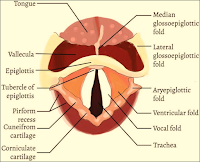
Larynx
➧ Despite the frequent use of tracheal intubation (TI) for both short-term events like surgery or for long-term ventilatory support, traumatic complications from TI still occur.

➧ Both the upper and lower airways are at risk of injury. The upper airway includes the naso- and oro-pharynx and extends to the level of the vocal cords. The lower airway lies distal to the vocal cords. 


1-Lips injury:

➧ Frequently the right lower lip is caught between the laryngoscope blade and the lower teeth. 

➧ It can also occur when the endotracheal tube (ETT) is placed or when a hard plastic oral airway is placed. 

➧ There is frequently soft tissue injury to the lip, but serious consequences are rare.

2-Dental injury:

➧ The upper incisors are usually involved. Identified risk factors include preexisting poor dentition and one or more indicators of difficult laryngoscopy and intubation. 

➧ A protective plastic shield placed over the upper teeth may limit the potential for damage to teeth.

3-Tongue injury:

➧ Prolonged compression by an ETT, an oral airway, or both may impair circulation, leading to ischemia and poor venous drainage, which then leads to macroglossia. 

➧ Obstruction of the submandibular duct by an ETT may lead to massive tongue swelling. 

➧ Compression injury to the lingual n. during difficult intubation has been reported with a one-month loss of tongue sensation.

4-Uvular injury:

➧ Injury to the uvula is a rare complication of TI and is usually associated with mechanical interference with the blood supply to the uvula during intubation. 

➧ Common causes of such interference include compression caused by excessive length of the ETT leading to direct pressure, blind pharyngeal suction with a hard suction catheter, and entrapment of the uvula between the ETT and oral airway. 

➧ This condition causes sore throat, difficulty in breathing, painful swallowing, and foreign body sensation in the throat. 

➧ Treatment of patients with steroids and antibiotics is successful, with complete healing in 2 weeks.

5-Nasal injury:

➧ Nasal intubations in particular are associated with mucosal tears or lacerations that may result in significant epistaxis. 

➧ The use of nasal decongestants/vasoconstrictors, lubrication of the ETT, and warming of the tube before nasal intubation may reduce the risk of epistaxis. 

➧ Once the nasotracheal tube is in place, it is important to position the tube in such a way as to prevent distortion of the nares, which can lead to local ischemia, followed by necrosis. 

➧ Blind nasal intubation also increases the risk of oropharyngeal and laryngeal injury, as does oral intubation with a protruding stylet. 

➧ Pharyngeal injury can progress into retropharyngeal infections or mediastinitis, and positive pressure ventilation may produce subcutaneous emphysema, pneumomediastinum, and pneumothorax.

6-Temporal-Mandibular Joint injury:

➧ This joint can be dislocated during TI.

7-Cervical Spine injury:

➧ Risk factors for spinal injury include head and neck trauma, cervical osteoporosis, atlantoaxial instability as seen in patients with rheumatoid arthritis and Down's syndrome, and lytic bone lesions. 

➧ Immobilization of the head and neck will prevent most spine injuries regardless of the route or method of intubation.

8-Pharyngeal and Esophageal injury:

➧ Venous drainage from the pharynx may be impaired by the mechanical presence of the tracheal tube and result in pharyngolaryngeal edema. 

➧ Both pharyngeal and esophageal perforations can occur after a difficult TI and in patients older than 60 y.

9-Laryngeal injury:

a) Vocal Cord Paralysis:

➧ Vocal cord paralysis is attributed to nerve injury or mechanical injury. 

➧ It may be unilateral or bilateral, bilateral injury is riskier and frequently requires emergency reintubation or tracheotomy. 

➧ The mechanism may be related to inflation of the ETT cuff at the level of the subglottic larynx rather than at the correct location in the trachea where an anterior branch of the recurrent laryngeal n. enters between the cricoid and the thyroid cartilage, providing innervation to the intrinsic muscles of the larynx. 

➧ An inflated cuff at this location can compress the nerve between the cuff and the overlying thyroid cartilage, causing injury. 

➧ This can be exacerbated when N₂O is used as part of the general anesthesia, as it diffuses rapidly into the cuff, increasing cuff pressure (Pcuff) and risk for injury. 

➧ Vocal cord immobility may also be caused by pseudolaryngeal paralysis which can be associated with cricoarytenoid dislocation, arytenoid fusion, and posterior glottic stenosis. 

➧ Interarytenoid fibrous adhesion can occur after intubation and is frequently confused with bilateral vocal cord paralysis. 

➧ Measures to decrease the risk for recurrent laryngeal n. injury include: 

-Use of a low-pressure, high-volume ETT cuff 

-Avoidance of larger than necessary ETTs 

-Avoidance of overinflation of the ETT cuff 

-Prevention of excessive tube migration during anesthesia 

➧ Vocal cord paralysis is usually associated with spontaneous recovery.

b) Arytenoid Cartilage Dislocation:

➧ The arytenoid cartilage can be dislocated from the cricoarytenoid joint due to the pressure of the tip of the tracheal tube as it is placed through the vocal cords. 

➧ This rare complication can occur with routine elective intubation and most often during traumatic intubation, intubations in which the patient is not relaxed and is struggling or coughing during tube placement and with the use of certain airway devices such as the McCoy laryngoscope or the lighted stylet (lightwand). 

➧ Acutely dislocated arytenoid cartilage can be reduced using rigid bronchoscopy. 

➧ If the dislocation is not reduced, chronic hoarseness and vocal cord dysfunction often result.

c) Hematoma and Granuloma formation:

➧ Hematoma formation on the vocal cords has been noted after routine TI and usually resolves without sequelae. 

➧ Occasionally scarring of the injured area occurs, resulting in a chronically hoarse voice. 

➧ Granulomas can also form after hematomas on the vocal cords resulting from TI. 

➧ Granuloma formation after intubation has been described as occurring with an incidence of 1:800 to 1:20000 in adults. 

➧ The most common site is the vocal process of the arytenoids because this structure comes into close contact with the ETT. 

➧ The degree of injury increases with increasing tube size and duration of intubation.

10-Tracheal injury:

➧ Tracheal laceration as a result of intubation was first reported in 1977. 

➧ Factors related to this type of injury include: 

-Overinflation of the ETT cuff 

-Multiple intubation attempts 

-Use of stylets 

-Malpositioning of the tube tip 

-Tube repositioning without cuff deflation 

-Inadequate tube size 

-Vigorous coughing 

-N₂O in the cuff 

-Traumatic intubation 

-Emergency intubation 

-Anesthesia protocol 

-An oversized tube 

-Long duration of intubation. 

➧ The risk is also greater in patients with: 

-Tracheal distortion caused by neoplasm or large lymph nodes 

-Weakness in the membranous trachea (seen in women or the elderly) 

-Chronic obstructive lung disease 

-Corticosteriod therapy. 

➧ The most common site for tracheal rupture is the junction of the cartilage and posterior membrane on the right side and the length of the tear often corresponds to the length of the cuff on the ETT. 

➧ Translaryngeal ETTs can cause circumferential stenosis or malacia at the cuff site. Tracheal stenosis can develop after even a brief (&lt;24 h.) TI. 

➧ The incidence of injury at the cuff site was likely higher in the era of high-pressure, low-volume cuffs and has certainly declined with the use of low-pressure, high-volume cuffs since the 1970s. 

➧ The newer cuffs, however, can easily be overinflated, exceeding the tracheal mucosal capillary perfusion pressure of 20-30 mmHg, and resulting in local tissue ischemia, which is directly proportional to the tracheal tube Pcuff. (Methods to stabilize ETT intracuff pressure)

➧ At a Pcuff of 30 cmH₂O, the tracheal mucosal blood flow becomes partially obstructed, and at a pressure of 45 cmH₂O, the obstruction becomes total, leading to tracheal mucosal damage and subsequent complications. 

➧ These lesions heal with either fibrous stenosis or loss of cartilaginous support and ensuing malacia.

Diagnosis:

➧ The clinical manifestations of laryngeal and tracheal injury can range from mild hoarseness to severe stridor. 

➧ Patients also may present with other non-specific symptoms such as exertional dyspnea and positional wheezing. 

➧ The airway is typically narrowed to less than 8 mm before exertional dyspnea is present. 

➧ Once the lumen is less than 5 mm, symptoms of dyspnea and stridor may present at rest. 

➧ Due to the dramatic loss of airway diameter before the development of symptoms, up to 54% of patients with tracheal stenosis can present in respiratory distress.

Treatment:

➧ Treatment of airway injury from prolonged intubation varies with the cause of injury but may include: 

-Bronchoscopy and dilation 

-Laser treatments of granulomas and webs 

-Resection of stenotic tracheal rings 

-Splitting the cicoid gland (cartilage).