Educational Blog about Anesthesia, Intensive care and Pain management

Fat Embolism Syndrome (FES)

Fat Embolism Syndrome (FES)

➧ Fat embolism can be difficult to diagnose. It most often follows a closed fracture of a long bone but there are many other causes.

Epidemiology:

➧ Incidence of this complication ranges from 0.5 to 11% in different studies. It varies considerably according to the cause. Patients with fractures involving the middle and proximal parts of the femoral shaft are more likely to experience fat embolism. Age also seems to be a factor with young men at the highest risk.

Etiology:

1-Fractures: Closed fractures produce more emboli than open fractures. Long bones, pelvis, and ribs cause more emboli. Sternum and clavicle cause less. Multiple fractures produce more emboli.

2- Orthopedic Procedures: Intramedullary nailing of long bones, Fixation of cemented hip prosthesis.

3-Massive soft tissue injury

4-Severe burns

5-Liposuction

6-Bone marrow biopsy

7-Bone marrow harvesting and transplant

8-Cardio-pulmonary bypass

9-Non-traumatic settings occasionally lead to fat embolism. These include conditions associated with: 

-Fatty liver

-Acute pancreatitis

-DM

-Osteomyelitis

-Bone tumor Lysis

-Pathological fractures

-Sickle cell crisis (causing bone infarcts)

-Decompression sickness

-Prolonged corticosteroids therapy

-Cyclosporine A solvent

-Parenteral lipid infusion

Sevitt’s classification:

1-Subclinical (Incomplete) form: Fat emboli present in blood and lungs, no symptoms/signs

2-Non-fulminant (Classic) form: Pulmonary dysfunction, cerebral dysfunction, petechiae

3-Fulminant form: Rare, rapid onset (within hours) of acute cor-pulmonale, respiratory failure, coma, and death.

Principal clinical features (triad) of FES:

1-Respiratory failure (dyspnea and hypoxia)

2-Cerebral dysfunction (confusion)

3-Petechiae

➧ Clinical features usually present between 24-72 h. after trauma (lucid interval). This is especially after fractures, when fat droplets act as emboli, becoming impacted in the pulmonary microvasculature and other microvascular beds, especially in the brain.

➧ Embolism begins rather slowly and attains a maximum in about 48 h.

➧ The initial symptoms are probably caused by mechanical occlusion of multiple blood vessels with fat globules that are too large to pass through the capillaries.

➧ The vascular occlusion in fat embolism is often temporary or incomplete as fat globules do not completely obstruct capillary blood flow because of their fluidity and deformability.

➧ The late presentation is thought to be a result of hydrolysis of the fat to more irritating free fatty acids which then migrate to other organs via the systemic circulation.

➧ It has also been suggested that paradoxical embolism occurs from shunting.

Clinical Presentation:

➧ There is usually a latent period of 24-72 h. between injury and onset.

➧ Symptoms: There is vague pain in the chest and shortness of breath.

➧ Signs: The onset is sudden, with:

-Restlessness

-Fever occurs, often at more than 38.3° C with a disproportionate tachycardia.

-Drowsiness with oliguria is almost pathognomonic.

➧ CNS signs: including a change in the level of consciousness, are common. They are usually non-specific and have the features of diffuse encephalopathy with acute confusion, stupor, coma, rigidity, or convulsions. Cerebral edema contributes to neurological deterioration.

Diagnostic Criteria:

A) Gurd's and Wilson’s Criteria:

-Diagnosis requires at least one sign from the major criteria and at least four signs from the minor criteria. (Table 1)


Gurd's and Wilson’s Criteria
Table 1: Gurd's and Wilson’s Criteria

B) Schönfeld’s Criteria:

-Diagnosis requires a score of more than 5. (Table 2)


Schönfeld’s Criteria
Table 2: Schönfeld’s Criteria

C) The clinical diagnosis is assured if all three of the following criteria are present within 72 h. after traumatic fracture:

1-Unexplained dyspnea, tachypnea, arterial hypoxia with cyanosis, and diffuse alveolar infiltrates on chest X-ray.

2-Unexplained signs of cerebral dysfunction, such as confusion, delirium, or coma.

3-Petechiae over the upper half of the body, conjunctiva, oral mucosa, and retina.

Differential Diagnosis:

➧ Dyspnea, hypoxia, and abnormal CXR: can occur with thrombo-embolism and pneumonia.

➧ Cerebral dysfunction: can occur with hypoxia or meningitis but the rash of meningococcal septicemia is all over and spreads rapidly. It can also occur as a late feature of head injury.

➧ Thrombotic Thrombocytopenic Purpura (TTP).

Investigations:

CBC: 

-Hematocrit: is decreased, occurs within 24-48 h., and is due to intra-alveolar hemorrhage. 

-Platelets: are decreased.

Serum Lipase: is increased, but this is not pathognomonic as it occurs in any bone trauma.

Serum Calcium: is decreased.

Cytological Examination: urine, blood, and sputum may detect fat globules that are either free or in macrophages. This test has low sensitivity and a negative result does not exclude fat embolism. 

Arterial Blood Gases: will show hypoxia, PaO₂ usually less than 60 mmHg, and hypocapnia. Continuous pulse oximeter monitoring may enable hypoxia from fat embolism to be detected in at-risk patients before it is clinically apparent.

ECG: Ischemia, Rt. Ventricular strain, Rt. Axis deviation, RBBB, Arrhythmia.

Chest X-ray: may show evenly distributed, fleck-like pulmonary shadows (Snow Storm appearance), increased pulmonary markings, and dilatation of the right side of the heart.

CT Head: (to rule out intracranial pathology).

CT Chest: (to rule out chest pathology).

Management:

A) Supportive

1-Ensuring good arterial oxygenation: High flow rate of oxygen is given to maintain the arterial oxygen tension in the normal range.

2-Restriction of fluid intake and Diuretics: to minimize fluid accumulation in the lungs so long as circulation is maintained.

3-Maintenance of intravascular volume: is important because shock can exacerbate the lung injury caused by FES. Albumin has been recommended for volume resuscitation in addition to the balanced electrolyte solution, because it not only restores blood volume but also binds fatty acids, and may decrease the extent of lung injury.

4-Mechanical ventilation (APRV) and Positive End-Expiratory Pressure (PEEP): may be required to maintain arterial oxygenation.

B) Pharmacological

1-Corticosteroids: Methylprednisolone (membrane stabilizer, decreases endothelial damage caused by free fatty acids).

2-Low dose heparin: 2500 u / 6 h. (reduce the degree of pulmonary comprise and intravascular coagulation despite the risk of hemorrhage and intravascular lipolysis).

3-Dextran-40: (decreases intravascular thrombosis when ESR is elevated).

4-Ethanol: (decreases lipolysis)

5-Dextrose: (decreases free fatty acid mobilization)

6-DVT prophylaxis

7-Nutrition

C) Surgical:

1-Prompt surgical stabilization of long bone fractures within 24 h. reduces the risk of the syndrome.

2-Use of vacuum or venting during reaming of long bones.

3-Prophylactic IVC filter in at-risk patients.

Prognosis:

➧ The mortality rate from FES is 5-15%. Even severe respiratory failure associated with fat embolism seldom leads to death.

➧ The prognosis is worse in older patients and those with more severe injury but is not affected by gender.

➧ Criteria of bad prognosis: Sevitt’s class, Serum lipase, Lipuria, ARDS.

Prevention:

➧ Early immobilization of fractures seems to be the most effective way of reducing the incidence of this condition.

Porphyric Crisis

Porphyric Crisis (Acute Neurovisceral Crisis) 

Background: 

-The porphyrias are caused by enzyme deficiencies in the heme production pathway. Such deficiencies may be due to inborn errors of metabolism or exposure to environmental toxins or infectious agents.

-The disease was named porphyria due to the red discoloration of urine in affected patients, (Figure 1).

-Crises are four to five times more common in women and usually occur in their early 30s.

Porphyria red urine
Figure 1: Porphyria Red Urine

Triggering factors: 

Enzyme-inducing drugs: 

-Barbiturates (Thiopental, Methohexital), Etomidate, Enflurane, Alcuronium, Mepivacaine, Pentazocine, Nifedipine, Verapamil, Diltiazem, Phenytoin, Hydralazine, Phenoxybenzamine, Aminophylline 

Physiological: 

-Menstruation, Fasting, Dehydration, Stress, Infection, Anemia, Endogenous hormones 

Habits: 

-Smoking, Alcohol 

Clinical Picture: 

CNS: 

-Autonomic neuropathy (Fever, Pain, Constipation, Gastroparesis, Postural hypotension) 

-Peripheral neuropathy (Skeletal muscle weakness, Quadriparesis, Bulbar palsy, Respiratory failure) 

-Cranial nerve palsy 

-Seizures 

-Psychiatric features (Mood disturbance, Confusion, Psychosis) 

CVS: 

-Tachycardia, Hypertension 

GIT: 

Acute abdominal pain, Vomiting, Diarrhea, Dehydration, Electrolyte disturbance (↓ [Na⁺, K⁺, Mg⁺²]) 

Management: 

Remove triggering factors (above) 

Specific R: 

-Hematin, Heme arginate / Heme albumin, Somatostatin, Plasmapheresis 

Symptomatic R: 

-Pain: Substantial doses of Opioids 

-Nausea and Vomiting: Prochlorperazine, Ondansetron 

-Anxiety: Lorazepam, Midazolam in low doses 

-Insomnia: Zopiclone 

-Delirium: Haloperidol 

-Tachycardia & Hypertension: β-adrenergic blocking agents, Glyceryl trinitrate 

-Seizures: Benzodiazepines, Levetiracetam, Clonazepam, Gabapentin, Vigabatrin, Magnesium sulphate 

-Sedation: Propofol, Alfentanil infusions. The clinical safety of prolonged midazolam infusion is unknown. 

-Thromboembolic prophylaxis: LMW heparins 

-Stress ulcer prevention: IV Omeprazole, Ranitidine 

-Correction of electrolytes

Mechanical Ventilation for Respiratory failure

Treacher Collins Syndrome

Treacher Collins Syndrome:


Treacher Collins Syndrome
-A craniofacial defect associated with developmental anomalies of the first arch. 

-Abnormalities vary from minimal, to complete syndrome. 

-The syndrome is named after Edward Treacher Collins, an English surgeon and ophthalmologist, who described its essential traits in 1900. 

-Patients may require anesthesia for maneuvers to improve upper airway obstruction temporarily, or for correction of some of the congenital defects. 

-Airway obstruction and a requirement for multiple operations increase the need for tracheostomy. 

Etiology:

-It is due to mutations in TCOF1, POLR1C, or POLR1D genes. TCOF1 gene mutations are the most common cause of the disorder, accounting for 81 to 93% of all cases. POLR1C and POLR1D gene mutations cause an additional 2% of cases. In individuals without an identified mutation in one of these genes, the genetic cause of the condition is unknown. 

Preoperative Abnormalities:

1. Features may include mandibular and malar hypoplasia, antimongoloid palpebral fissure, macrostomia, irregular maloccluded teeth, microphthalmia, lower lid defects, cleft palate, high arched palate, macroglossia, and auricular deformities. 

2. Associated abnormalities include mental retardation, deafness, dwarfism, cardiac defects, choanal atresia, and skeletal deformities. 

3. Chronic upper respiratory tract obstruction and obstructive sleep apnea, which can lead to growth retardation and/or cor-pulmonale. 

Anesthetic Problems:

1. Upper airway obstruction: in neonates, this may require urgent temporary maneuvers, such as stitching the tongue to the lower lip 

2. Excess secretions: may impede induction of anesthesia 

3. Inhalational induction: may be difficult 

4. Difficult tracheal intubation 

5. Obstructive sleep apnea may occur postoperatively 

6. Pulmonary edema 

Intraoperative Management:

Recommendations:

1-Monitoring by pulse oximetry, to detect airway obstruction, is crucial. 

2-Avoid respiratory depressant drugs, both for premedication and postoperatively 

3-Use drying agents 

4-Never give muscle relaxant until the airway has been secured. 

Airway Management:

-Several methods have been proposed to overcome the problem of difficult intubation, some in the awake patient and some under general anesthesia: 

a) In Awake Patients: 

1-Awake intubation or awake direct laryngoscopy to visualize the vocal cords. 

2-Direct laryngoscopy, with the patient in the sitting position, and using a 5-G feeding tube taped to the side of the laryngoscope to give oxygen. 

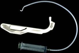
3-The Augustine guide (Figure 1): can be used for nasotracheal intubation in an awake, sedated patient.

Augustine guide
Figure 1: Augustine guide

4-Fibreoptic bronchoscope or Tracheostomy under local anesthesia: 

In small infants, the ‘tube over bronchoscope’ technique is not always possible because of the small size of the tube, therefore a Seldinger-type approach may be necessary: 

-After the administration of atropine, ketamine IM, and topical lidocaine, a fibreoptic bronchoscope (OD 3.6mm, L 60 cm, and suction channel 1.2 mm) was passed through one nostril. 

-The tongue was held forward with Magill forceps, until the vocal cords were seen, but not entered, because of the risk of total obstruction. 

-Under direct vision, a Teflon-coated guidewire with a flexible tip was passed via the suction channel into the trachea. 

-The bronchoscope was carefully removed leaving the wire in place, and an ID 3mm nasotracheal tube was then passed over it into the trachea. 

Pediatric bronchoscopes of 2.5 mm diameter are now available, but their very fineness makes them less easy to handle than the 4-mm bronchoscopes). 

b) In Anesthetized Patients: 

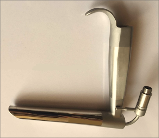
1-Jackson anterior commissure laryngoscope (Figure 2): 

-The head is elevated above the shoulders, with flexion of the lower cervical vertebrae and extension at the atlanto-occipital joint. The laryngoscope is introduced into the right side of the mouth. Only the tip is directed towards the midline, the proximal end remaining laterally so that a further 30 degrees of anterior angulation can be obtained. 

-The narrow, closed blade prevents the tongue from falling in and obscuring the view of the larynx. When visualized, the epiglottis is elevated, and the larynx is entered. Intubation is then achieved by passing a lubricated tube, without its adaptor, down the laryngoscope. It is held in place with alligator forceps whilst the laryngoscope is withdrawn.

Jackson anterior commissure laryngoscope
Figure 2: Jackson anterior commissure laryngoscope

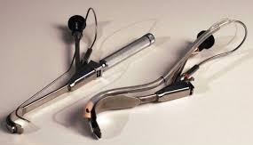
2-The Bullard intubating laryngoscope (Figure 3):

-It can be used to achieve nasotracheal intubation.

Bullard intubating laryngoscope
Figure 3: Bullard intubating laryngoscope

3-Tactile nasal intubation: 

-Inhalational induction with sevoflurane, and the tongue is pulled downwards and forwards. The tube is initially used as a nasal airway, whilst the index and middle finger are used to palpate the epiglottis, through which the tube is then passed. 

4-Laryngeal mask airway: 

-Inserted under propofol anesthesia and can be used as a conduit for the passage of a fibreoptic bronchoscope.

5-The use of an assistant to pull out the tongue with Magill forceps, and at the same time to apply cricoid pressure, to assist laryngoscopy. 

Postoperative Management:

-The tracheal tube should remain in place until the patient is fully awake. 

-Patients should be nursed in a high-dependency area postoperatively. The combination of sleep apnea and drugs with CNS-depressant effects may make them particularly susceptible to respiratory arrest.

Read more ☛ Pierre Robin Syndrome

Bone Cement Implantation Syndrome (BCIS)

Bone Cement Implantation Syndrome (BCIS)

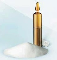
Components of Bone Cement

Bone Cement constituents:

-Bone cement is an acrylic polymer that is formed by mixing two sterile components: 

a) Powder:

1-Polymer: Polymethyl methacrylate/co-polymer (PMMA) 

2-Initiator: Benzoyl peroxide (BPO) 

b) Liquid:

1-Monomer: Methyl methacrylate (MMA) 

2-Accelerator: N, N-Dimethyl para-toluidine (DMPT)/diMethyl para-toluidine (DMpt) 

-To make the cement radiopaque, a contrast agent is added, either zirconium dioxide (ZrO₂) or barium sulphate (BaSO₄). 

-When the two components are mixed, the liquid monomer polymerizes around the pre-polymerized powder particles to form hardened PMMA. During this polymerization process, heat is generated, due to an exothermic reaction, and reaches temperatures of around 82–86 °C. 

Incidence:

-The incidence of BCIS in cemented orthopedic procedures is approximately 20%. 

-The incidence of a severe reaction resulting in cardiovascular collapse within this group is 0.5-1.7%. 

Orthopedic procedures incidence:

-Cemented hemiarthroplasty (highest incidence) 

-Total hip replacement 

-Knee replacement surgery 

BCIS typically occurs during:

-Bone cementation and prosthesis insertion 

-Femoral reaming (before cementation) 

-Joint reduction and limb tourniquet deflation (after cementation) 

Patients at high risk of cardio-respiratory compromise:

-Male sex 

-Increasing age 

-ASA class III / IV 

-Significant cardiopulmonary disease 

-Chronic obstructive pulmonary disease 

-Use of diuretics 

-Use of warfarin 

Conditions that can increase the incidence of BCIS:

-Osteoporosis, Bone metastasis, and Concomitant hip fractures. 

These conditions may be associated with increased or abnormal vascular channels, through which marrow contents can more readily migrate into the circulation, resulting in emboli. 

Pathophysiology of BCIS:

a) Embolization of the Medullary Contents:

-During surgical cementation and prosthesis insertion, the cement is intentionally pressurized to force it into the interstices of the bone, to improve bonding between the cement and bone. 

-The cement then expands in the space between the bone and the prosthesis, further pressurizing air and the medullary contents, forcing them into circulation. These embolic contents include fat, marrow, cement, air, bone particles, and aggregates of platelets and fibrin. 

-When these medullary contents embolize, they may reach the lungs, heart, and/or coronary circulation, causing the characteristic hypoxia and right ventricular dysfunction, leading to hypotension. 

b) Histamine release, Hypersensitivity, and Complement activation:

-Contact with bone cement (Methyl Methacrylate), leads to an increase in blood levels of anaphylactoid complements (C3a and C5a) and histamine, which are potent mediators of vasoconstriction and bronchoconstriction. 

-These mediators result in an increase in pulmonary vascular resistance, causing ventilation/perfusion disturbances, hypoxia, right ventricular failure, and cardiogenic shock. 

Clinical Picture:

-Hypoxia 

-Sudden loss of arterial blood pressure 

-Pulmonary hypertension 

-Arrhythmias 

-Loss of consciousness 

-Cardiac arrest 

-Under general anesthesia, a significant drop in systolic blood pressure (SBP) may herald cardiovascular collapse, whilst a sudden drop in end-tidal pCO₂ may indicate right heart failure leading to a catastrophic reduction in cardiac output. 

-In an awake patient under a regional anesthetic, early signs of BCIS may include dyspnea and/or altered sensorium. 

BCIS Severity Spectrum:

a) Non-fulminant BCIS:

-Characterized by a significant, yet transient, reduction in arterial oxygen saturation and SBP in the peri-cementation period. 

b) Fulminant BCIS:

-With profound intraoperative cardiovascular changes, progressing to; arrhythmias, shock, or cardiac arrest. 

Classification of BCIS Severity: based on the degree of hypoxia, hypotension, and conscious level (Table 1)

Classification of BCIS Severity
Table 1: Classification of BCIS Severity

Prophylaxis:

a) Anesthesia:

1-Ensure adequate hemodynamic optimization pre- and intra-operatively 

2-Keep SBP within 20% of the pre-induction value 

3-Prepare vasopressors in case of cardiovascular collapse 

4-Confirm awareness that cement is about to be prepared/applied 

5-Maintain vigilance for cardiorespiratory compromise 

b) Orthopedic:

1-Inform the anesthetist before cement application 

2-Wash and dry the femoral canal 

3-Apply cement retrograde, utilizing a suction catheter and intramedullary plug in the femoral shaft 

4-Avoid excessive pressurization (3rd generation technique) 

Management:

-Administration of 100% inspired oxygen is first-line therapy, with airway control if necessary. 

-Invasive hemodynamic monitoring (if not already in place), should be established. 

-In cases of severe BCIS (when the patient has been arrested, or in a peri-arrest condition), standard advanced cardiopulmonary life support (ACLS) algorithms and procedures should be followed. 

-Fluid resuscitation to maintain Rt. Ventricle preload, and inotropes to support ventricular contractility.

-Vasopressors (such as Phenylephrine and Noradrenaline) primarily cause peripheral vasoconstriction, and increase aortic blood pressure, which in turn supports coronary artery blood flow, and thus improves myocardial perfusion and contractility. 

-Use of vasopressors and inotropes should be continued into the postoperative period as necessary, under the management of the intensive care unit (ICU).